Areas of Research
Basic Neuroscience and Brain Imaging Methods

The role of preference in the emotional benefits of nature exposure
Functional near infrared spectroscopy (fNIRS) is a neuroimaging technique that measures the same biological signal as fMRI and can be used in a variety of less restrictive settings than an MRI scanner. In this study, participants completed an N-back working memory task while fNIRS data were recorded. As expected, participants showed greater fronto-parietal cortical activity during a more difficult 2-back task vs. an easier 1-back task. However, the results for the 3-back task were less consistent and it was hypothesized that this was due to somewhat low accuracy on the 3-back task. To see how task accuracy might be influencing the results, a behavioral partial least squares (PLS) analysis was conducted, which is a data-driven multivariate technique that has been used in other neuroimaging modalities to identify brain-behavior associations. Results of this analysis showed that the relationship between fNIRS activity (defined by change in deoxyhemoglobin concentrations (HbR) in mid-frontal cortex) and task accuracy does indeed differ depending on task difficulty (N-back level). More specifically, a larger reduction in HbR (equivalent to increased neural activity) was positively correlated with higher performance on the 3-back task, unrelated to activity on the 2-back task, and negatively correlated with performance on the 1-back task.
Stable long-range interhemispheric coordination is supported by direct anatomical projections
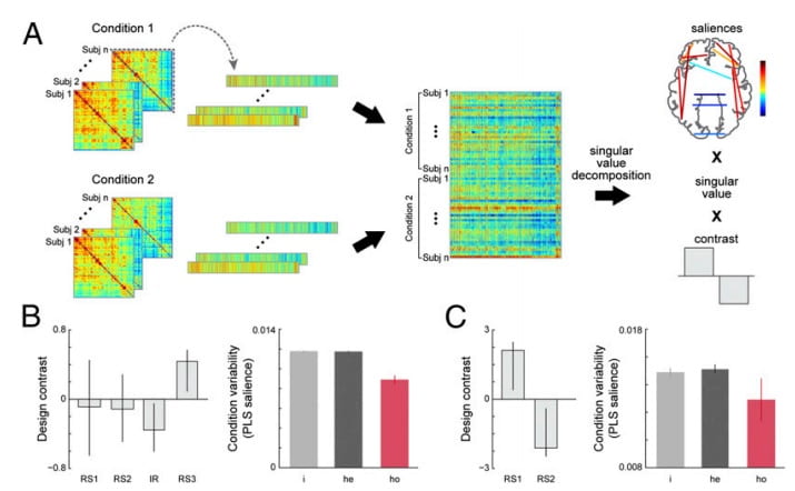
We show that the functional coordination between the two hemispheres of the brain is maintained by strong and stable interactions of a specific subset of connections between homotopic regions. Our data suggest that the stability of those functional interactions is mediated in part by the direct anatomical projections of large, highly myelinated fibers that traverse the corpus callosum. These functional properties were evident in both humans and macaques, suggesting a preserved framework for interhemispheric communication despite an increase in functional lateralization in humans. These results contribute to our fundamental understanding of how dynamic functional interactions between the two hemispheres of the mammalian brain are supported by its underlying anatomical architecture.
The Functional Connectivity Landscape of the Human Brain
Functional brain networks emerge and dissipate over a primarily static anatomical foundation. The dynamic basis of these networks is inter-regional communication involving local and distal regions. It is assumed that inter-regional distances play a pivotal role in modulating network dynamics. Using three different neuroimaging modalities, 6 datasets were evaluated to determine whether experimental manipulations asymmetrically affect functional relationships based on the distance between brain regions in human participants. Contrary to previous assumptions, here we show that short- and long-range connections are equally likely to strengthen or weaken in response to task demands. Additionally, connections between homotopic areas are the most stable and less likely to change compared to any other type of connection. Our results point to a functional connectivity landscape characterized by fluid transitions between local specialization and global integration. This ability to mediate functional properties irrespective of spatial distance may engender a diverse repertoire of cognitive processes when faced with a dynamic environment.


Pattern Classification of fMRI data: Applications for analysis of spatially distributed cortical networks
The field of fMRI data analysis is rapidly growing in sophistication, particularly in the domain of multivariate pattern classification. However, the interaction between the properties of the analytical model and the parameters of the BOLD signal (e.g. signal magnitude, temporal variance and functional connectivity) is still an open problem. We addressed this problem by evaluating a set of pattern classification algorithms on simulated and experimental block-design fMRI data. The set of classifiers consisted of linear and quadratic discriminants, linear support vector machine, and linear and nonlinear Gaussian naive Bayes classifiers. For linear discriminant, we used two methods of regularization: principal component analysis, and ridge regularization. The classifiers were used (1) to classify the volumes according to the behavioral task that was performed by the subject, and (2) to construct spatial maps that indicated the relative contribution of each voxel to classification. Our evaluation metrics were: (1) accuracy of out-of-sample classification and (2) reproducibility of spatial maps. In simulated data sets, we performed an additional evaluation of spatial maps with ROC analysis. We varied the magnitude, temporal variance and connectivity of simulated fMRI signal and identified the optimal classifier for each simulated environment. Overall, the best performers were linear and quadratic discriminants (operating on principal components of the data matrix) and, in some rare situations, a nonlinear Gaussian naïve Bayes classifier. The results from the simulated data were supported by within-subject analysis of experimental fMRI data, collected in a study of aging. This is the first study that systematically characterizes interactions between analysis model and signal parameters (such as magnitude, variance and correlation) on the performance of pattern classifiers for fMRI.
Evaluating Functional Localizers: The Case of the FFA.
Functional localizers are routinely used in neuroimaging studies to test hypotheses about the function of specific brain areas. The specific tasks and stimuli used to localize particular regions vary widely from study to study even when the same cortical region is targeted. Thus, it is important to ask whether task and stimulus changes lead to differences in localization or whether localization procedures are largely immune to differences in tasks and contrasting stimuli. We present two experiments and a literature review that explore whether face localizer tasks yield differential localization in the fusiform gyrus as a function of task and contrasting stimuli. We tested standard localization tasks–passive viewing, 1-back, and 2-back memory tests–and did not find differences in localization based on task. We did, however, find differences in the extent, strength and patterns/reliabilities of the activation in the fusiform gyrus based on comparison stimuli (faces vs. houses compared to faces vs. scrambled stimuli).
