
Superresolution Microscopy
Sub-diffraction fluorescence imaging Check Microscope StatusNew User TrainingHands-on Microscopes
To access a microscope, click the New User Training button above and work through our training checklist. Trained users have 24/7 access to the facility and are given permission to schedule their own microscope sessions through our online scheduling software.
Stimulated Emission Depletion (STED)
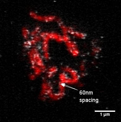
STED micrograph courtesy of V. Cloud, Doug Bishop lab
What is STED-CW? Stimulated Emission Depletion (STED) is an advanced form of laser scanning confocal microscopy that allows for a 3-4 fold improvement in resolution over traditional laser scanning confocal; from a smallest resolvable feature size of 150-200nm FWHM to approximately 50nm FWHM. This allows us to see objects that were previously below the limit of optical resolution for light microscopy (Abbe diffraction limit). CW (continuous wave) technology means that the depletion laser is continuous, not pulsed as in previous STED microscopes. This allows for superresolution at higher speeds than previously possible. See the Leica website for more information and example images.
For best results in STED-CW imaging: we recommend placing samples on a #1.5 coverslip (0.170mm thickness) mounted with ProLong Gold (Invitrogen) antifade mounting medium or Thiodiethanol (TDE). Ideally, samples should contain high quantum yield, photostable fluorophores such as: AlexaFluor 450/488/514, Dylight 488, Atto 425/488, eGFP, mCitrine, mVenus, or mCerulean. For information on STED imaging, or to set up a training session, contact Christine Labno or Vytas Bindokas.
Leica SP8 Laser Scanning Confocal with White Light Laser, 3D STED and FLIM
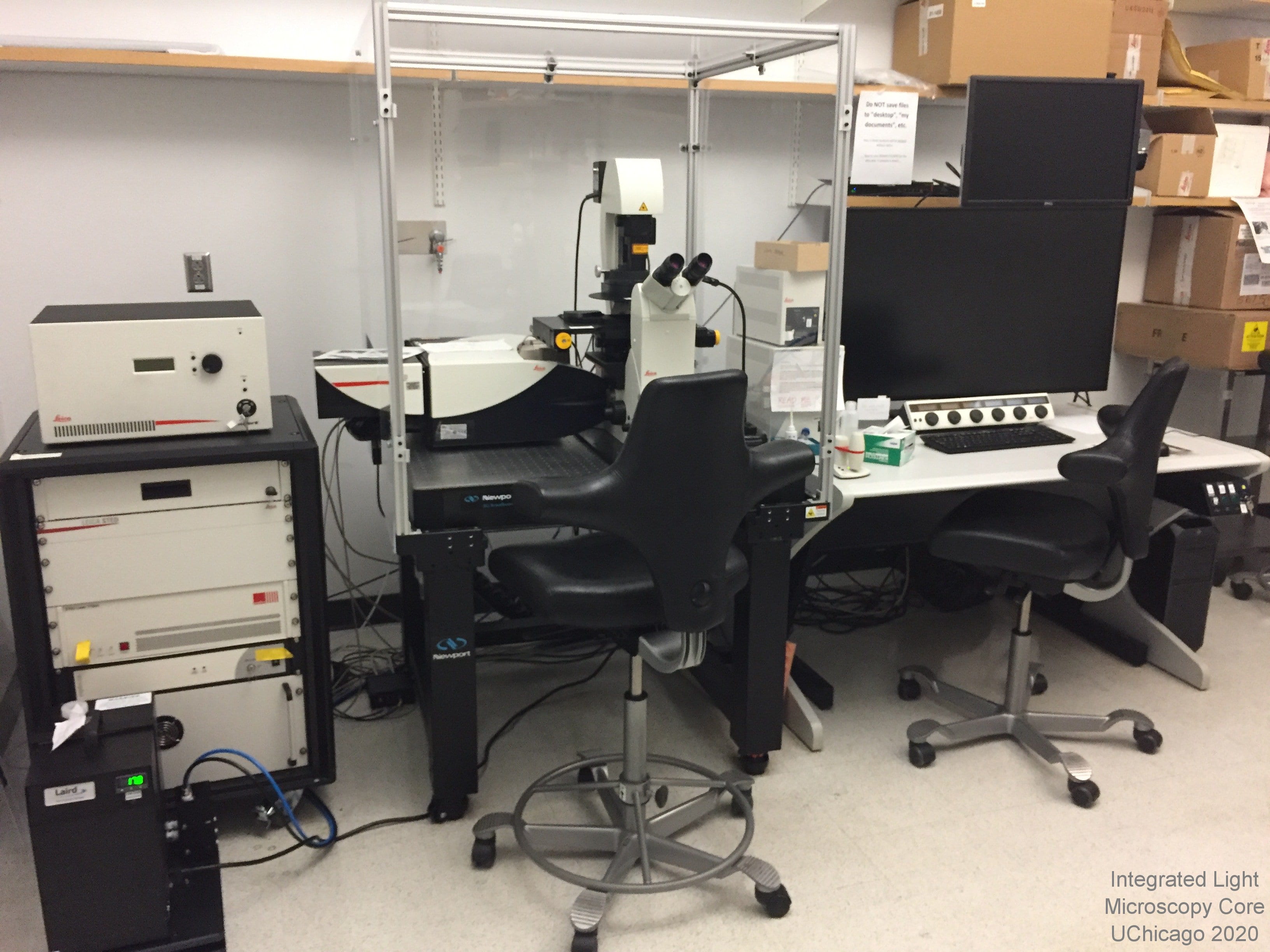 Overview: The Leica SP8 is a laser scanning confocal capable of fluorescence lifetime (FLIM), 3-color 3D STED and tau-STED imaging.
It features a white light laser (WLL) capable of excitation in any wavelength from 470nm to 670nm, and an acousto-optical beam splitter (AOBS) to select/introduce up to eight laser lines (at 8nm intervals) at a time. In addition to the WLL, there are Argon and UV lasers for imaging, photobleacing and photoactivation, so exciation spans the spectrum from UV to deep red. There are also three depletion lines for STimulated Emission Depeletion (STED) superresoluion: 775nm (depletes red and far red dyes), 660nm (depletes orange and yellow dyes) and 592nm (depletes green dyes). The acousto-optical tunable filters (AOTF) make it possible to detect a wide range of emission wavelengths with unlimited range on each of three HyDs (hybrid GaAsP detector) two PMTs (photon multiplier tube). Spectral scanning / unmixing in exciation and/or emission is available. Time gated fluorescence detection is possible on the three HyD detectors.
Location: KCBD 1250G
Training Contact: Lorraine Horwitz
Fluorophores this microscope can image:
- Violet/near UV (ex: DAPI, Alexa 405)
- Cyan (ex: CFP)
- Green (ex: GFP, Alexa 488)
- Yellow (ex: mCitrine, YFP)
- Orange (ex: Cy3, Alexa 543)
- Near Red (ex: Texas Red, Alexa 594)
- Far Red (ex: Cy5, Alexa 647)
- Near IR (ex: Alexa 700)
Excitation Light Source(s):
- White light laser -- ANY wavelength from 470nm - 670nm. Up to 8 lines at once with 8nm spacing
- Near UV laser at 405nm
- 5 line Argon laser at 458, 476, 488, 496 and 514nm
Lasers for STED depletion:
- pulsed 775nm (depletes red and far red dyes)
- continuous wave 660nm (depletes orange and yellow dyes)
- continuous wave 592nm (depletes green dyes)
Emission Detection:
- Custom emission detection of any wavelength range between 410nm and 800nm
- 2 chilled photon multiplier tubes (PMTs)
- 2 chilled, single-molecule sensitive hybrid GaAsP/PMT detectors (SMD-HyDs) with optional time gating
- 1 non-SMD hybrid GaAsP/PMT detector (HyD) with optional time gating
- Transmitted light detector
- 8-, 12-, or 16-bit grayscale output
Objectives (base magnification, additional optical zoom is possible):
- 10x / NA 0.4 dry wd=2.74mm 506407
- 20x / 0.7 multi-immersion (water, oil or glycerol) wd=0.66mm 506343
- 40x / 1.25 oil wd=0.24mm 506358
- 63x / 1.4 UV oil wd=0.14mm 506350
- 100x / 1.45 oil (STED-rated) wd=0.13mm 506378
- 93x / NA 1.3 glycerol wd=0.3mm 506417 (must be installed prior to use, STED-rated, motorized correction collar)
- 86x / NA 1.20 water wd=0.3mm 506333 (must be installed prior to use, STED-rated, motorized correction collar)
Sample Chamber:
- Inverted platform for imaging of slides or dishes
- Automated XY stage for tiling / multipoint scanning
- "SuperZ" galvo focusing stage, 1.5 mm range. Demonstrated capability of three cell volumes per second (12 x 1-mm optical slices each)
- Navigator software for non-rectangular tiling. Increases speed for large tiling projects. Includes a fast "stage overview" mode
- Chamber slide users see our chamber slide use warning before starting your cultures!
Special Features:
- Capable of Fluorescence Lifetime Imaging (FLIM) and lifetime enhanced STED (tau-STED) imaging
- Capable of 2D or 3D superresolution imaging in XY, XZ or XYZ.
- AOBS (acousto-optical beam splitter) plus sequential scanning capability allows for rapid sequential scanning of fluorophores with minimal bleed-through or cross-talk
- Hybrid GaAsP detectors (HyDs) and chilled SMD HyD detectors feature digital time gating (0.1 nanosecond increments, 3.5 nanosec. (min) to 12 nanosec (max) range) and photon counting mode.
- Transmitted light detector features DIC polarizer/analyzer with prisms for 20x, 40x and 63x.
- Notch filters for 488, 561, 594 and 633 from the WLL
- Spectral scanning across excitation, emission, or both, allowing for separation of fluorophores with similar ranges (e.g. GFP and FITC) through spectral unmixing.
- Time-gating on HyDs allows for differentiation of fluorophores with similar spectra or enhancement of STED resolution from the pulsed 775nm laser.
- Tandem scanner with dual scanning galvanometer mirrors allows for either high speed scanning (max 16,000 Hz scan rate) 25 images/sec at 512x512; strip scans to 333 fps (5 channels) OR high pixel density scanning (imaging to 4k x 4k [16 megapixels] per channel in galvo mode).
- Standard galvo scanner includes beam park for FRAP, bleaching and photoactivaiton
- Optical zoom for sampling to 5nm pixel size
- Wizards for FRAP, FRET and Live Data Mode
- 3D/4D reconstruction software built in to LAS_X image collection software
- LAS_X Leica confocal software on Windows 10
- Off-line version of LAS_X software available on facility workstation
Leica SP5 STED Laser Scanning Confocal
 NOTE: The 592 depletion laser on this system is no longer functional. We do not have a service contract for this end-of-life system, so we will not be replacing this laser. Anyone in need of confocal imaging with DAPI can use the SP5 2-photon, SP8 or Stellaris. Anyone in need of super-resolution imaging has likely already moved to the SP8 3 color 3D STED system. The rest of the system continues to function, so confocal with green, red and far red fluorophores is still possible.
Leica SP5 STED - October 18, 2023 - We swapped this Argon (488nm) laser with the one from the SP5 2-photon which was starting to malfunction. We recommend keeping the power level between standby and 20% (under configurations->laser) to decrease the chances of the laser malfunction and prolong the life of the laser. We do not know how long this laser will last, but we do know if the malfunction becomes more serious we will have to retire this microscope (see no service contract for this end-of-life system above).
Overview: The SP5 II is an advanced, high speed laser scanning confocal platform. It includes an acousto-optical beam splitter (AOBS) to select/introduce most excitation laser lines. There are eight excitation lines available, spanning the spectrum from green to far red. The acousto-optical tunable filters (AOTF) make it possible to detect a wide range of emission wavelengths with unlimited range on each of three PMTs (photo multiplier tubes), two HyDs (hybrid GaAsP detector) or two APDs (avalanche photodiodes).
Location: KCBD 1250F
Training Contact: Lorraine Horwitz
Fluorophores this microscope can image:
- Cyan (ex: CFP)
- Green (ex: GFP, Alexa 488)
- Yellow (ex: mCitrine, YFP)
- Orange (ex: Cy3, Alexa 543)
- Near Red (ex: Texas Red, Alexa 594)
- Far Red (ex: Cy5, Alexa 647)
- Near IR (possible but not ideal, ex: Alexa 700)
Excitation Light Source(s):
- 5 line Argon laser at 458, 476, 488, 496, and 514nm
- DPSS laser at 561nm
- orange HeNe laser at 594nm
- red HeNe laser at 633nm
Emission Detection:
- Custom emission detection of any wavelength range between 410nm and 800nm
- 3 chilled photon multiplier tubes (PMTs)
- 2 hybrid GaAsP/PMT detectors (HyDs) with optional time gating
- 2 internal avalanche photodiode detectors (APDs) for high sensitivity (green/red and CFP/YFP filters available)
- Transmitted light detector with DIC polarizer/analyzer plus prisms for most objectives available
- 8-, 12-, or 16-bit grayscale output
Objectives (base magnification, additional optical zoom is possible):
- 10x / NA 0.4 dry wd=2.74mm
- 20x / 0.7 multi-immersion (water, oil or glycerol) wd=0.26-0.17mm
- 40x / 1.25-0.75 oil wd=0.22mm
- 63x / 1.4-0.6 UV oil wd=0.14mm
- 100x / 1.40 oil (STED-rated) wd=0.13mm
- 50x / 0.9 dry wd=0.28mm (must be installed prior to use)
- 63x / 1.3 glycerol CORR wd=0.30mm (must be installed prior to use)
Sample Chamber:
- Full wrap incubator box with warm air heating for live samples
- Inverted platform for imaging of slides or dishes
- Automated XY stage for tiling / multipoint scanning
- "SuperZ" galvo focusing stage, 1.5 mm range. Demonstrated capability of three cell volumes per second (12 x 1-mm optical slices each)
- Chamber slide users see our chamber slide use warning before starting your cultures!
Special Features:
- Continuous wave depletion laser at 592nm allows for single or dual color super resolution (STED method) with either the resonant (high speed) or galvo (high pixel density) scanners.
- STED mode allows for resolution of particles down to 50nm FWHM (cyan, green, yellow fluorophores only)
- Tandem scanner with dual scanning galvanometer mirrors allows for either high speed scanning (max 16,000 Hz scan rate) 25 images/sec at 512x512; strip scans to 333 fps (5 channels) OR high pixel density scanning (imaging to 8k x 8k [64 megapixels] per channel).
- Standard scanner includes beam park for FRAP, bleaching and photoactivaiton
- Sequential scanning capability allows for rapid sequential scanning of fluorophores with minimal bleed-through or cross-talk
- Wizards for FRAP and FRET
- AOTF (acousto-optical tunable filters) for spectral scanning, allowing separation of fluorophores with similar ranges (e.g. GFP and FITC) through spectral unmixing
- LAS_AF Leica confocal software on Windows
- Off-line version of LAS_AF software available on facility workstation
Ground State Depletion (GSD)

Depth-coded GSD image courtesy of Jiping Yue, Xiaoyang Wu lab
What is GSD? Ground State Depletion is one of the “pointilist” techniques for superresolution microscopy. This group also includes Stochastic Optical Reconstruction Microscopy (STORM) and Photoactivated Localization Microscopy (PALM).”Pointilist” superresolution microscopy techniques use various methods to manipulate a fluorescently labeled sample into returning only a handful of diffraction limited (200nm) fluorophore signals at one time. These signals are then mathematically processed to localize them to sub-diffraction (20nm) spots. Thousands of frames worth of these localized spots are collected over a potentially minutes-long imaging time and integrated on the fly to form a complete image. Up to three fluorophores (green, red, deep red) can be imaged serially to create multi-color images. TIRF illuminated, 2D epifluorescent and 3D superresolution imaging are all possible.
Leica GSD/TIRFM Ground State Depletion Superresolution Microscope
The Leica GSD was provided by the Institute for Genomics & Systems Biology (IGSB) and the Institute for Molecular Engineering (IME).
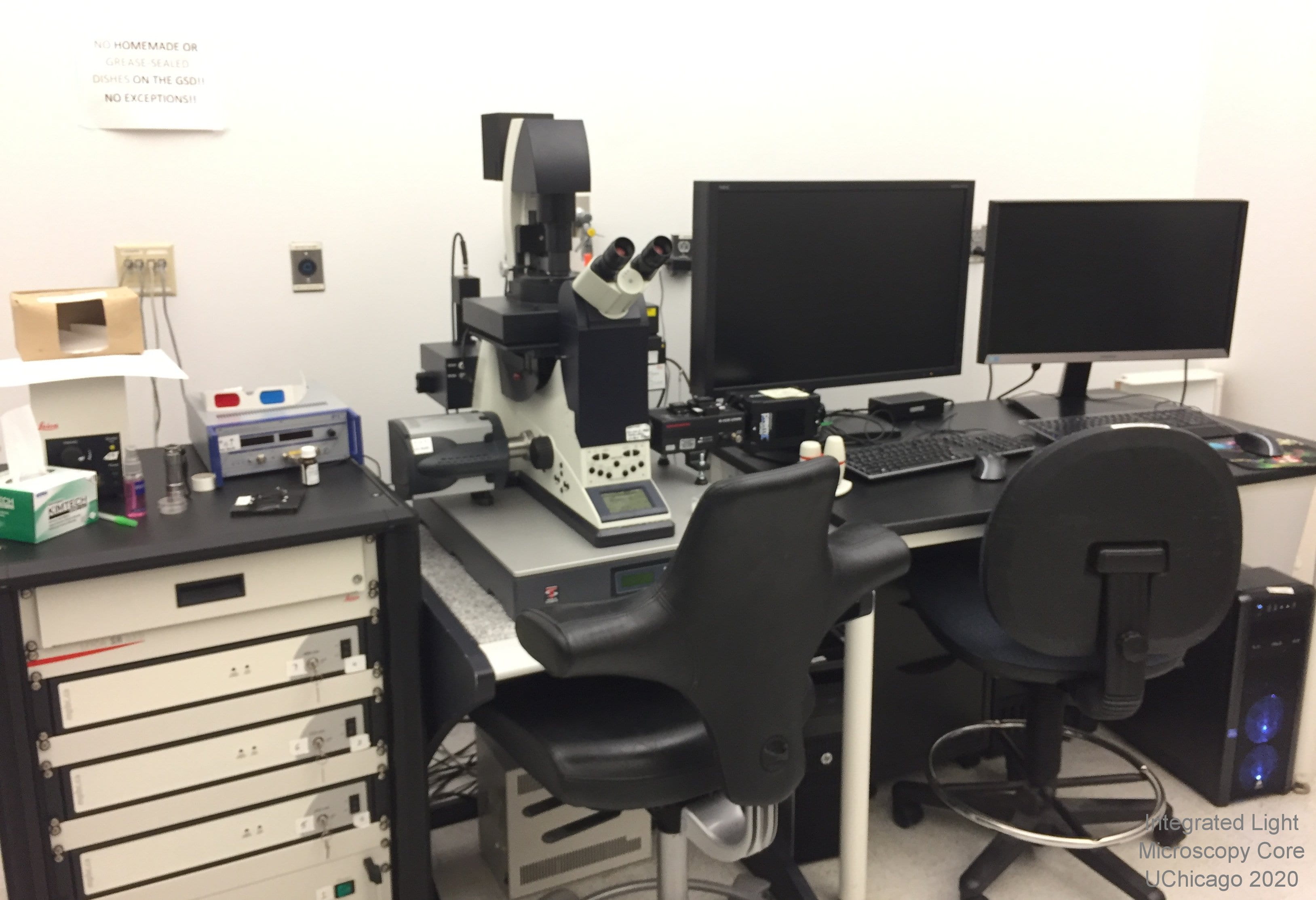 Overview:The Leica GSD is a four-color Total Internal Reflection fluorescence (TIRF) and a three-color Ground State Depletion (GSD) microscope. It is not a confocal microscope. There are four excitation lines available, spanning the spectrum from violet to far red. All can be used for TIRF; green to far red can be used for GSD.
Location: KCBD 1250B
Training Contact: Christine Labno
Fluoropohores this microscope can image:
- Blue (ex: DAPI, Alexa 405) - TIRFM ONLY, NO GSD
- Green (ex: GFP, Alexa 488)
- Orange (ex: Cy3, Alexa 543)
- Near Red (ex: Texas Red, Alexa 594)
- Far Red (ex: Cy5, Alexa 647)
Excitation Light Source(s):
- solid state violet laser at 405nm (used to "backpump" which increases fluorophore blinking, can also be used for TIRF excitation)
- solid state blue laser at 488nm
- green fiber laser at 532nm
- red fiber laser at 642nm
Excitation and Emission Filters:
- Green: simultaneous excitation at 400-410 and 483-493nm with dichroic at 496nm and emission at 425-475nm and 505-605nm
- OPTIONAL manual green band pass filter for emission at 503-547nm
- Red: simultaneous excitation at 400-410nm and 527-537nm with dichroic at 541nm and emission at 425-475nm and 550-650nm
- OPTIONAL manual red band pass filter for emission at 582-636nm
- Far Red: simultaneous excitation at 400-410nm and 637-647nm with dichroic at 649nm and emission at 425-475nm and 660-760nm
- OPTIONAL manual long pass filter for far red emission at 664nm (664 LP)
- Quad: ex 400-410nm; 483-493nm; 27-537nm; 637-647nm dichroics: 417, 496, 544, 655 em: 421-477nm; 497-519nm; 547-621nm; 666-732nm
Emission Detection:
- iXon Ultra EMCCD camera for high frame rate, low light imaging with low read noise
- Option to use a Photometrics Prime95B camera and Hamamatsu w-Gemini image splitter operated by a second imaging computer. This allows simultaneous two-color GSD acquisition and/or development of biplane 3D localization
Objectives:
- 160x / 1.43 oil 0.07mm WD - this is a state-of-the-art, adhesive-free objective developed by Leica exclusively for GSD imaging
- 10x, 20x, 40x and 60x DRY objectives are available for ocular viewing and widefield images ONLY
Sample Chamber:
- Inverted platform
- Small laser safety chamber for imaging on #1.5 coverslips or 35mm glass-bottom dishes (sorry, NO heated stage or CO2 at this time)
- Manual XY stage movement
- PiFoc precision focusing / Z step control
Special Features:
- Supressed Motion (SuMo) stage which locks the 160x objective to the stage to minimize sample drift
- Camera-based, software-driven TIRF laser focusing and alignment.
- Software-based TIRF angle setting allows for your choice of multiple laser angles, creating varying evanescent wave penetration depths. TIRF imaging direction (north, south, east or west) can also be selected
- Two off-line workstations for data processing
Proper Sample Preparation for Ground State Depletion: Samples should be mounted or grown on #1.5 thickness coverslips (high tolerance Schott glass if possible) and stained with high quantum yield fluorophores such as: AlexaFluor 647, 555, or 488, Atto 647 or 488, Rhodamine 6G, Cy5, Cy3b or bodipy (this is not an exhaustive list). DO NOT use DAPI! Also, DO NOT mount the coverslips to slides. Bring unmounted coverslips in the holding medium of your choice (PBS works well) for imaging. We will provide MEA mounting buffer and apply it just before imaging. For more information on GSD sample prep contact Christine Labno or Vytas Bindokas.
Super-Resolution by Optical Re-assignment (SoRa)
SoRa 3i Marianas Spinning Disk Confocal
Overview: Fully automated, inverted, Yokogawa-type spinning disk confocal ideal for imaging live samples. Features an automated XY stage and piezo-controlled fine Z stage (focus) positioning for mutlipoint scanning plus incubator box for temperature, CO2 and humidity control. Slidebook software controls filter cube turret, objective turret, condenser, DIC and brightfield optics and high-speed brightfield shutter. SoRa with microvolution for nearly instantaneous deconvolution is ideal for super-resolution live cell imaging with little phototoxicity.
Location: KCBD 1250F
Training Contact: Khalil Rodriguez
Fluorophores this microscope can image:
- Violet/near UV (ex: DAPI, Alexa 405)
- Cyan (ex: CFP)
- Green (ex: GFP, Alexa 488)
- Yellow (ex: mCitrine, YFP)
- Orange (ex: Cy3, Alexa 543)
- Near Red (ex: Texas Red, Alexa 594)
- Far Red (ex: Cy5, Alexa 647)
Excitation Light Source(s):
- Near UV laser at 405nm
- Solid state laser at 445nm
- Solid state laser at 488nm
- Solid state laser at 561nm
- Solid state laser at 638nm
Emission Detection:
- Two Prime 95B Back Illuminated Scientific CMOS (11x11um square pixels @ 18.7mm field of view)
- band pass filter for blue
- band pass filter for cyan
- band pass filter for green
- band pass filter for red
- band pass filter for far red
Objectives:
- 10x / NA 0.3 dry (Plan-Neofluar) working distance: 5.2mm
- 40x / NA 1.3 oil (Plan-Apochromat)
- working distance: 0.22mm
- 63x / NA 1.4 oil (Plan-Apochromat)
- working distance: 0.14mm
- 100x / NA 1.46 oil (Alpha Plan-Fluar)
- working distance: 0.11mm
Sample Chamber:
- Inverted platform for imaging of slides or dishes
- OKO full environmental control chamber (constant temperature, humidity and CO2)
- Motorized XY stage for multipoint timelapse and tiling
- Piezo controlled fine Z-stage positioning for 3D imaging
Special Features:
- Super-Resolution through Optical Reassignmnet (SoRa) is ideal for super-resolution live cell imaging and low phototoxicity
- CSU-W1 Yokogawa spinning disk allows for high speed imaging up to 200fps, wide field of view 16mm x 17mm, 25µm and 50µm pinhole disks for lower and higher magnification objectives
- Microvolution GPU accelerated Deconvolution Module for nearly instantaneous deconvolution.
- Options for split-view imaging, NIR imaging, illumination field flattening and super-resolution imaging
- Fast shutter speeds and channel switiching for high speed imaging
- Vector high-speed point scanner for photoactivation/photoablation and FRAP experiments
- Full microscope automation through Slidebook software
Structured Illumination Microscopy (SIM)
Lattice Lightsheet Bessel Beam Illumination Microscope
 Overview:This is a commerically-produced clone of the Betzig system described in Science October 2014 and currently running at the HHMI Janelia Campus’s Advanced Imaging Center. It is designed for low-light 3D time lapse imaging. This is NOT like the Zeiss Lightsheet.Z1 meant for large preps! Imaging is by means of water-immersion objectives and is limited to less than 100 micrometers depth of penetration.
Sample Preparation: All samples MUST be on 5mm round coverslips. Use of this microscope requires a Staff person to operate the system. Please be sure to arrange your session in advance with Core Staff. Contact Vytas Bindokas or Christine Labno (information at left).
Special Features:
- High speed, low light 3D timelapse imaging.
- Heated bath chamber for live cell work. Perfusion is possible but we have no CO2 at this time
- Hamamatsu Flash4v2+ sCMOS high-sensitivity, low noise camera
- 100nm pixel size (xy)
- Nikon 25x NA 1.1 water immersion objective
- 405nm (DAPI), 488nm (GFP), 561nm (mCherry) and 642nm (Cy5) lasers
- LED for brightfield illumination