
Hands-on Microscopes
To access a microscope, click the New User Training button above and work through our training checklist. Trained users have 24/7 access to the facility and are given permission to schedule their own microscope sessions through our online scheduling software.
Total Internal Reflection Fluorescence Microscopy (TIRFM)
What is TIRFM? Total Internal Reflection Fluorescence Microscopy is used to observe events near the plasma membrane of cells, or the fluorescence of a single molecule, without interference from fluorescence coming from the cell’s cytoplasm. An evanescent wave selectively excites fluorophores in a small region (less than 200 nm) above the coverglass. It is ideally suited to processes such as insulin granule release, cell / matrix adhesion, and 2D protein trafficking
Leica GSD/TIRFM Ground State Depletion Superresolution Microscope
The Leica GSD was provided by the Institute for Genomics & Systems Biology (IGSB) and the Institute for Molecular Engineering (IME).
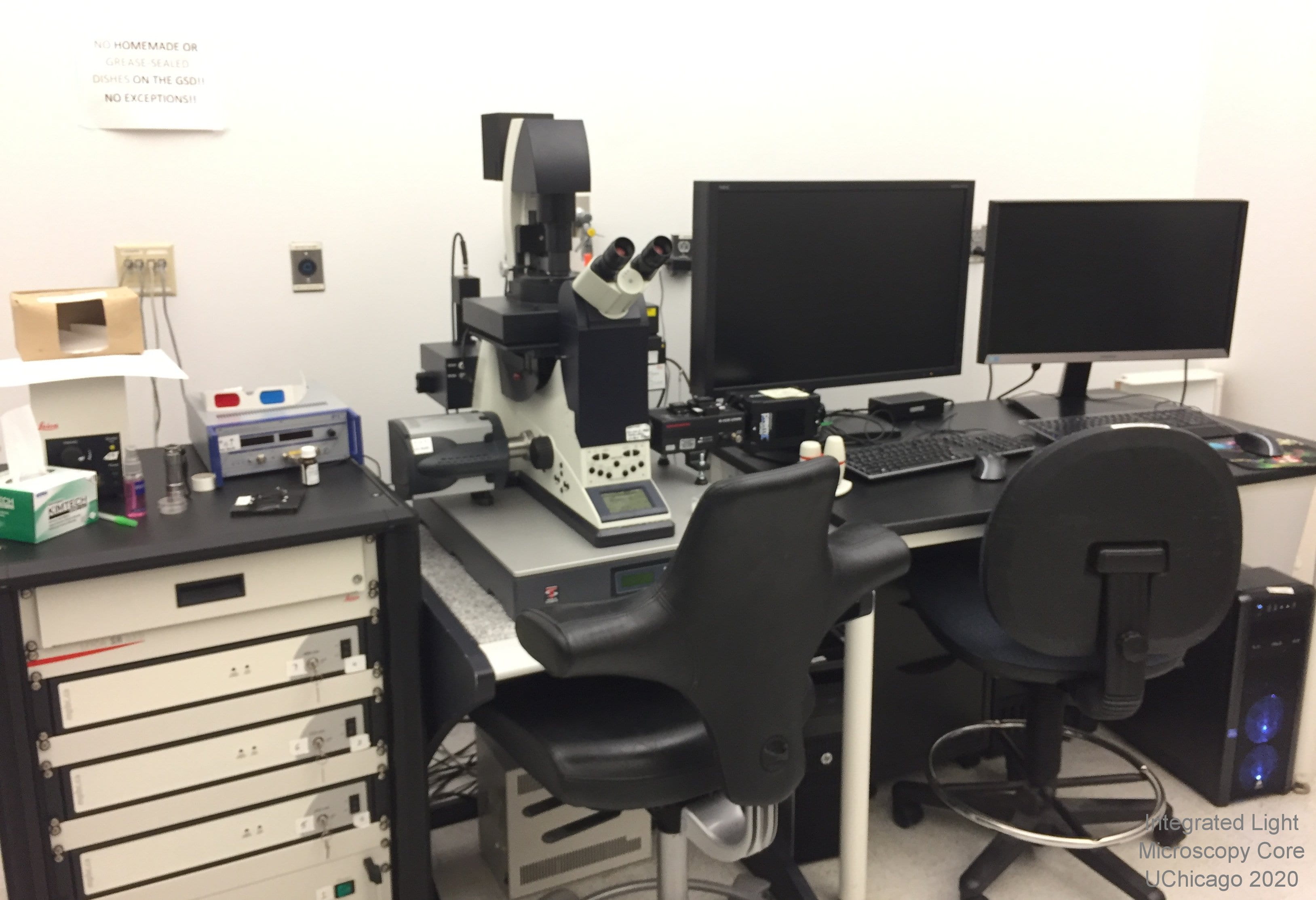 Overview:The Leica GSD is a four-color Total Internal Reflection fluorescence (TIRF) and a three-color Ground State Depletion (GSD) microscope. It is not a confocal microscope. There are four excitation lines available, spanning the spectrum from violet to far red. All can be used for TIRF; green to far red can be used for GSD.
Location: KCBD 1250B
Training Contact: Christine Labno
Fluoropohores this microscope can image:
- Blue (ex: DAPI, Alexa 405) - TIRFM ONLY, NO GSD
- Green (ex: GFP, Alexa 488)
- Orange (ex: Cy3, Alexa 543)
- Near Red (ex: Texas Red, Alexa 594)
- Far Red (ex: Cy5, Alexa 647)
Excitation Light Source(s):
- solid state violet laser at 405nm (used to "backpump" which increases fluorophore blinking, can also be used for TIRF excitation)
- solid state blue laser at 488nm
- green fiber laser at 532nm
- red fiber laser at 642nm
Excitation and Emission Filters:
- Green: simultaneous excitation at 400-410 and 483-493nm with dichroic at 496nm and emission at 425-475nm and 505-605nm
- OPTIONAL manual green band pass filter for emission at 503-547nm
- Red: simultaneous excitation at 400-410nm and 527-537nm with dichroic at 541nm and emission at 425-475nm and 550-650nm
- OPTIONAL manual red band pass filter for emission at 582-636nm
- Far Red: simultaneous excitation at 400-410nm and 637-647nm with dichroic at 649nm and emission at 425-475nm and 660-760nm
- OPTIONAL manual long pass filter for far red emission at 664nm (664 LP)
- Quad: ex 400-410nm; 483-493nm; 27-537nm; 637-647nm dichroics: 417, 496, 544, 655 em: 421-477nm; 497-519nm; 547-621nm; 666-732nm
Emission Detection:
- iXon Ultra EMCCD camera for high frame rate, low light imaging with low read noise
- Option to use a Photometrics Prime95B camera and Hamamatsu w-Gemini image splitter operated by a second imaging computer. This allows simultaneous two-color GSD acquisition and/or development of biplane 3D localization
Objectives:
- 160x / 1.43 oil 0.07mm WD - this is a state-of-the-art, adhesive-free objective developed by Leica exclusively for GSD imaging
- 10x, 20x, 40x and 60x DRY objectives are available for ocular viewing and widefield images ONLY
Sample Chamber:
- Inverted platform
- Small laser safety chamber for imaging on #1.5 coverslips or 35mm glass-bottom dishes (sorry, NO heated stage or CO2 at this time)
- Manual XY stage movement
- PiFoc precision focusing / Z step control
Special Features:
- Supressed Motion (SuMo) stage which locks the 160x objective to the stage to minimize sample drift
- Camera-based, software-driven TIRF laser focusing and alignment.
- Software-based TIRF angle setting allows for your choice of multiple laser angles, creating varying evanescent wave penetration depths. TIRF imaging direction (north, south, east or west) can also be selected
- Two off-line workstations for data processing
Proper Sample Preparation for Ground State Depletion: Samples should be mounted or grown on #1.5 thickness coverslips (high tolerance Schott glass if possible) and stained with high quantum yield fluorophores such as: AlexaFluor 647, 555, or 488, Atto 647 or 488, Rhodamine 6G, Cy5, Cy3b or bodipy (this is not an exhaustive list). DO NOT use DAPI! Also, DO NOT mount the coverslips to slides. Bring unmounted coverslips in the holding medium of your choice (PBS works well) for imaging. We will provide MEA mounting buffer and apply it just before imaging. For more information on GSD sample prep contact Christine Labno or Vytas Bindokas.