
Hands-on Microscopes
To access a microscope, click the New User Training button above and work through our training checklist. Trained users have 24/7 access to the facility and are given permission to schedule their own microscope sessions through our online scheduling software.
Zeiss AxioObserver 7 automated, inverted widefield

Overview: Fully automated AxioObserver 7 widefield microscope with incubation chamber, fluorescence and DIC optics, full color histology, Prior motorized XY stage, and high NA objectives with auto-immersion capabilities at a wide range of magnifications and excellent Axiocam 705 monochrome and Axiocam 305 color CMOS cameras. Zeiss Zen software (blue edition) with AI automated sample finder, XYZT automation including time lapse, slide scanning, and mark and find multipoint over time capabilities. Fluorescence filters for DAPI, cyan, green, yellow, red, far red, and near IR plus DIC prisms for select objectives.
Location: KCBD 1250F
Training Contact: Lorraine Horwitz
Fluorophores this microscope can image:
- blue (e.g. DAPI)
- cyan (e.g. CFP)
- green (e.g. GFP/Alexa 488)
- yellow (e.g. YFP)
- red (e.g. DsRed, mCherry, Alexa 594)
- far red (e.g. Cy5, Alexa 647)
- near IR (e.g. cy7)
Excitation Light Source:
- LED light source
Emission Detection:
- Monochrome and color CMOS cameras
Objectives:
- 5x dry
- 10x dry
- 20x dry
- 40x / 1.1 water - rewatering feature
- 63x / 1.4 oil
Sample Chamber: XYZ automated inverted microscope stage
Olympus IX81 inverted widefield microscope
 Overview: This is an inverted, automated fluorescence microscope with a motorized stage and full-wrap incubation chamber. It is well suited to multi-well plate imaging, fixed cell fluorescence imaging, live cell imaging with real-time pixel intensity readouts, FRET experiments, ratiometric imaging (such as fura-2 340/380), and checking transfection efficiency.
Location: KCBD 1250C
Training Contact: Christine Labno
Fluorophores this microscope can image:
- Blue (e.g. DAPI)
- Cyan (e.g. CFP)
- Green (e.g. GFP, Alexa 488) -- 2 filter sets
- Yellow (e.g. YFP, mCitrine)
- Red (e.g. mCherry, Alexa 543) -- 2 filter sets
- Far red (e.g. Cy5, Alexa 647) -- 2 filter sets
- Near IR (e.g. Cy7)
- Fura-2 ratiometric Ca++ dye
Excitation and Emission filters:
- penta (5 color) dichroic with narrow filters / fast switching - blue ex: 387/11 em: 440/40 dichroic: 408; greens ex: 485/20 HQ em: 525/32 dichroic: 504; reds ex: 560/25 em: 650/13 dichroic: 581; far reds ex: 607/36 em: 684/24 dichroic 667; near IR ex: 740/13 em: 809/81 dichroic 762
- CFP ex: 436/20 and em: 480/40 (must be installed prior to use)
- green single cube - ex: 480/40 HQ and em: 510LP
- YFP ex: 500/20 and em: 535/30HQ (must be installed prior to use)
- red single cube - ex: 530-550 and em: 590LP
- far reds (Cy5) single cube - ex: 620/60 HQ and em: 700/75 HQ
- Fura-2 ex: 340; 380 and em: 510/40
- Chameleons 2 ex: 440/20 and em: 535/35; 485/40 (must be installed prior to use)
Excitation Light Source: Mercury arclamp
Emission Detection: Hamamatsu Orca Flash 4.0 camera with 4 megapixels (6.5um pixel size, 2048 x 2048 pixels). Huge field of view, great for tissue samples and multi-well plates
Objectives:
- 4x / 0.16 dry
- 10x / NA 0.3 dry (UPlanFL)
- 20x / 0.4 dry (LC PlanFL)
- 40x / 1.35 oil UV (UApo/340, best for FURA)
- 60x / 1.45 oil (PlanApo, TIRF rated)
- 100x 1.45 oil (TIRF rated)
- Also available (must be swapped in):
- 2x / 0.06 dry
- 40x / 0.6 dry LWD
- 150x / 1.45 oil (TIRF rated)
Sample Chamber:
- Automated, inverted platform (model IX81) for imaging on slides, live cell dishes or multi-well plates
- Full-wrap incubator box with micro CO2 chamber available
- Marzhauzer xy motorized stage for precise control of tiling, multi-point sampling or multi-well plate reading
- Chamber slide users see our chamber slide use warning before starting your cultures!
Special Features:
- SlideBook imaging software image capture in 2D, 3D, time lapse, multipoint, tiling, multi-well or any combination
- Zero Drift Correction (TM) auto re-focusing system. Especially useful for long term time lapse and / or heated preps
- DIC polarizer/analyzer plus appropriate prisms for most objectives
- Fura-2 340/380 ratiometric imaging
- CFP/YFP and Chameleon FRET filter set
- Automated filter changer with single color filters and a penta dichoic / filters for fast multi-color fluorescence imaging
- A beam splitter is available, but not currently installed. Be aware there is significant vignetting due to the large chip size of the Flash 4.0 camera. The beam splitter is for simultaneous capture of two emission wavelengths with the SAME excitation wavelength. Splitters available are: green/red (520/30 and 630/50) and blue/yellow-orange (460/50 and 570/60).
Olympus SZX-Zb12 Stereomicroscope
 Overview: The stereomicroscope is built for imaging large specimens. The scope has fluorescent, transmitted, and oblique illumination, 2 objectives, zoom focusing, and a high resolution color camera.
Location: KCBD 1250C
Training Contact: Christine Labno
Fluorophores this microscope can image:
- Blue (e.g. DAPI)
- Green (e.g. GFP, Alexa 488)
- Red (e.g. mCherry, Alexa 594)
Excitation Light Sources:
- Stereo fluorescence with 100W mercury lamp
- External fiber optic illuminator for oblique white lighting
Emission Detection:
- Hamamatsu Orca Flash 4.0 camera with 4 megapixels (6.5um pixel size, 2048 x 2048 pixels). Huge field of view, great for tissue samples and multi-well plates
- Zeiss Axiocam 14-bit CCD digital color camera (6.7um pixel size,1300x1035 pixels)
Objectives:
- 0.5x plus zoom 3.5-45x
- 1.2x plus zoom 8.4-108x
Special Features:
- Large field of view
- 10x occulars
- Long working distance and wide zoom range (12:86:1)
- Slidebook 6.0 software for fluorescence and AxioVision SE64 Rel. 4.8 software for full color histology
Zeiss Axiovert 200M inverted widefield microscope - DECOMMISSIONED NOV 2023
This microscope is no longer available for use, it has been replaced with a Zeiss Axio Observer 7. The information here is for publication generation purposes only.
Click for user's guide
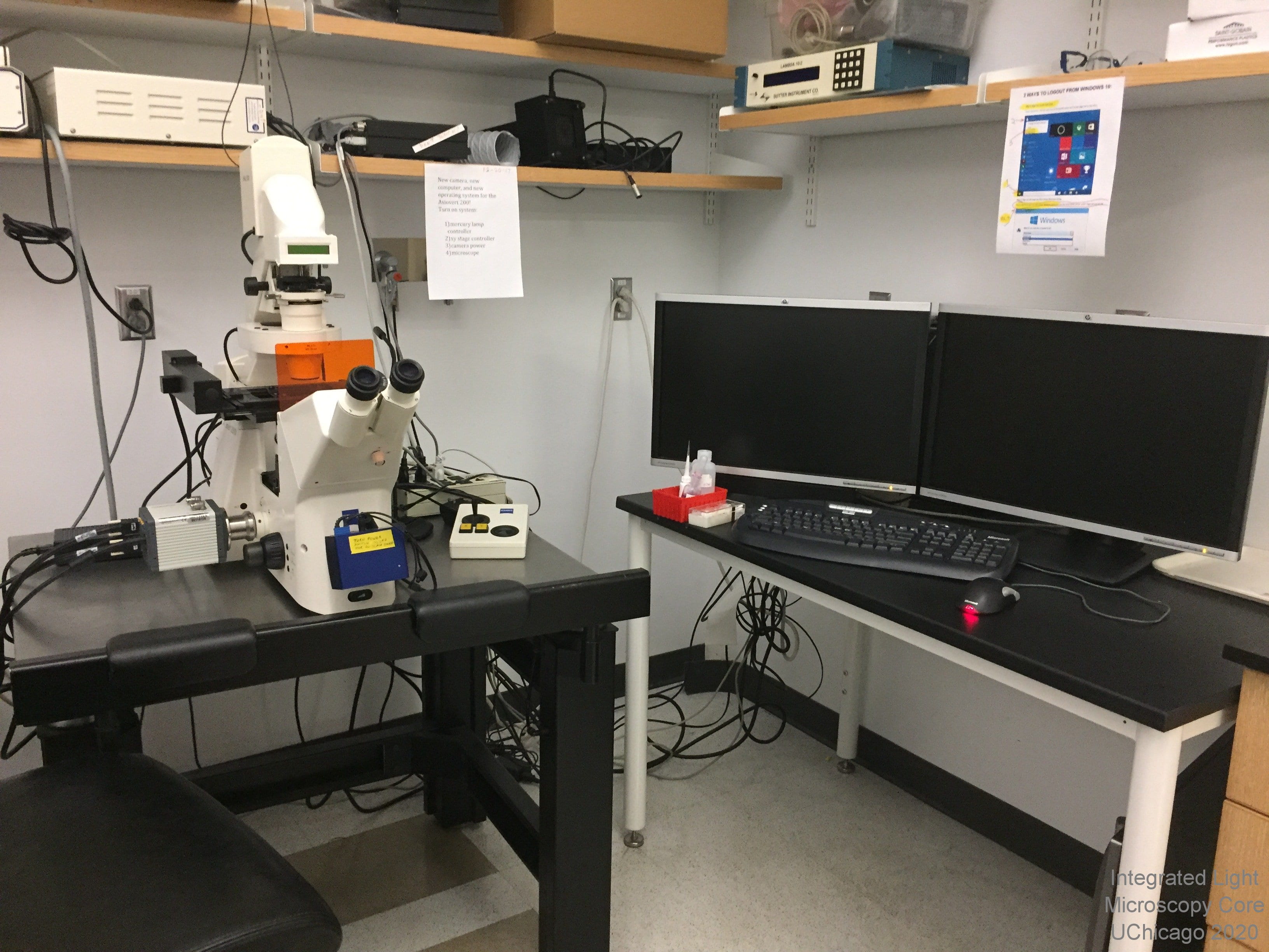 Overview: The Axiovert 200M is a fully motorized, inverted widefield microscope from Zeiss. This microscope is used for immunofluorescence, Nomarksi (DIC) or full color histology images on fixed or live specimens on slides. The high quality images can be further enhanced by digital deconvolution on the analysis stations. Users may capture their images using the ORCA ER digital camera for fluorescent samples or the Zeiss Axiocam color digital camera for samples which require transmitted light color imaging.
Location: This microscope is no longer available for use
Training Contact: N/A
Fluorophores this microscope can image:
- Blue (e.g. DAPI)
- Green (e.g. GFP, Alexa 488)
- Red (e.g. mCherry, Alexa 594)
- Far Red (e.g. Alexa 647)
- Full color histology imaging with white light
Emission Detection:
- Hamamatsu Orca Flash 4.0 camera with 4 megapixels (6.5um pixel size, 2048 x 2048 pixels). Huge field of view, great for tissue samples and multi-well plates
- Zeiss Axiocam 14-bit CCD digital color camera (6.7um pixel size,1300x1035 pixels)
Objectives:
- 10x / 0.5 NA
- 20x / 0.5 NA
- 40x / 1.3 NA oil
- 40x Korr
- 63x / 1.4 NA oil
Sample Chamber:
- Inverted platform and multiple stages to support slides and dishes for live and fixed samples
- Automated z-drive for z-stacks
- Automated xy stage for fine movement
- Chamber slide users see our chamber slide use warning before starting your cultures!
Special Features:
- SlideBook software for image capture
- Automated reflector turret with 5 positions and a multitude of fluorescent filters
- Automated condenser turret
- Dual filter wheels with individual excitation filters for fluorescence
- Nomarksi (DIC) imaging
Olympus VivaView in-incubator microscope - DECOMMISSIONED FEB 2024
 This microscope is no longer available for use, it has been replaced with a Zeiss Axio Observer 7. The information here is for publication generation purposes only.
Overview: The Olympus LCV110U VivaView is an innovative system for long-term, multi-dimentional, live cell imaging. The system is an inverted widefield fluorescence microscope built into an incubator box. The incubator keeps optimal temperature, humidity and CO2 during long experiments and decreases the effects of thermal drift and evaporation. Allows for imaging of multiple areas in up to eight dishes, kept at 37C, 5% CO2. Brightfield, blue (DAPI), cyan (CFP), green (GFP) and near red (mCherry, DsRed) imaging available.
Location: This microscope is no longer available for use
Training Contact: N/A
Fluorophores this microscope can image:
- blue (e.g. DAPI)
- cyan (e.g. CFP)
- greens (e.g GFP, Alexa 488)
- reds (e.g. mCherry, Alexa 543)
- far reds (e.g. Cy5, Alexa 633)
Excitation Light Source and Filters:
- X-Cite eXactate arclamp light source with liquid light guide can be adjusted to lower light intensity and minimize photobleaching.
- blue excitation filter at 387/11nm; dichroic 409nm
- cyan excitation filter at 436/20; dichroic 445
- green excitation filter at 470-495; dichroic 505
- red excitation filter at 530-550; dichroic 570
- far red excitation filter at 620/60; dichroic 700
Emission Detection and Filters:
- Air-cooled (-35C) CCD camera gives 12-bit grayscale images with 1344 x 1024 pixels (6.45 micron pixels)
- blue emission filter at 447/60
- cyan emission filter at 480/40
- green emission filter at 510-550
- red emission filter at 575IF (long pass)
- far red emission filter at 700/75
Objective:
- 40x / NA 0.95 dry
- 0.5x and 2x magnification changers create 20x and 80x magnification
Sample Chamber:
- Fully enclosed, humidified incubator set to 37C, 5% CO2.
- Inverted platform with 8 position motorized carousel for imaging multiple areas across multiple 35mm round live-cell dishes (#1.5 glass bottom dishes recommended, MatTek dishes are available in sleeves of 10 in the facility)
Special Features:
- Independent objective and carousel heaters minimize thermal drift
- MetaMorph image capture software
- Data are saved to hard drive immediately post-capture
- Optional motorized z-stack collection for 3D or focus correction
Drop Off Service
We have two slide scanners that are run purely as a drop-off service.
To use the service: first see our Slide Preparation and Practical Considerations documents.
First-time Core users will need to make sure they have access to their lab's OSRF FTP server account and submit a Facility User Record.
All users need to bring a completed copy of the Scanner Service Request Form and your slides in a container labeled with your name to KCBD 1250
NOTE ON CLINICAL SAMPLES - All slides containing patient samples must be de-identified before scanning (i.e. protected health information must be covered, blacked out or otherwise removed)
We will add your slides to the queue and email you when your scans are done. Scan files will be available for download on the OSRF FTP server. Physical slides should be picked up from designated areas as soon as possible after scanning. Slides not picked up within 3 months become the property of the Core and we will discard them if we run out of storage space.
To view your images: For CRi .mrxs files use CaseViewer (PC only) or QuPath; for Olympus VS200 .vsi files use OlyVIA or QuPath. We have created guides for basic OlyVIA and basic QuPath use
Contact Khalil Rodriguez for more information on this service
Olympus VS200 Slideview Research Slide Scanner
Click for freeOlyVIA software download or free QuPath whole slide image analysis software from the University of Edinburgh
The Core has created a guide for beginning OlyVIA software use (in progress) and a guide for beginning QuPath use with whole slide scans. The QuPath developers have extensive documentation as well.
Click for our Slide Preparation Guidelines and our Practical Considerations for Slide Scanning
Overview: Slide scanning on the Olympus VS200 Slideview is run as a drop off service. Our whole slide scanners create a virtual library of histology or multi-channel fluorescent slides. Details on the virtual slide can then be zoomed to a virtual magnifications from 0.5x-100x and exported as .tif or .jpeg for analysis or presentation with other software packages. We currently recommend processing with either the opensource QuPath software package or the proprietary free download from Olympus called OlyVIA.
Location: KCBD 1250E
Drop off Contact: Khalil Rodriguez
This microscope can image:
- Full color histology imaging with white light
- Blue (DAPI) ex 378/52 em 432/36 (Quad dichroic)
- Cyan (CFP) ex 438/24 em 482/25 (Triple dichroic)
- Green (FITC, GFP) ex 474/27 em 515/30 (Quad dichroic)
- Yellow (YFP) ex 509/22 em 544/24 (Triple dichroic)
- Orange (TRITC) ex 554/23 em 595/31 (Quad dichroic)
- Red (mCherry) ex 578/21 em 641/75 (Triple dichroic)
- Far red (Cy5) ex 635/18 em 698/70 (Quad dichroic)
- Near IR (Cy7) ex 735/28 em 809/100 (Single dichroic)
Excitation Light Sources:
- X-Cite Xylis broad spectrum LED from Encelitas
Emission Detection:
- Hamamatsu Orca Fusion digital camera for fluorescence (3.45um pixel size binned 2x2 for final 6.9um pixel size, 5.3 megapixels with 80% QE)
- Integrated 2/3 inch CMOS for histology (3.45 μm × 3.45 μm pixel size, high sensitivity, high resolution)
Objectives:
- 2x, 4x, 10x NA 0.4, 20x NA 0.8, and 40x NA0.95 dry
- 60x NA 1.42 oil (small regions only, file size and scan time can balloon quickly)
Pixel Sizes:
- 2x - 3.275 um/pixel (fluorescence), 2.7382 um/pixel (histology)
- 4x- 1.507 um/pixel (fluorescence), 1.3691 um/pixel (histology)
- 10x - 0.6504 um/pixel (fluorescence), 0.548 um/pixel (histology)
- 20x- 0.3251 um/pixel (fluorescence), 0.274 um/pixel (histology)
- 40x - 0.1626 um/pixel (fluorescence), 0.137 um/pixel (histology)
- 60x - 0.1084 um/pixel (fluorescence), 0.091 um/pixel (histology)
Special Features:
- Drop off service: drop off your slides and put your name in the queue. We'll do the rest and notify you when your scans are done. Slides are scanned on a first-come, first-served basis.
- Automated scanning of up to 210 slides per run, unlimited slides per batch
- Data is available on our server, so you can download it straight to any campus computer.
- Offline image viewing software available free from Olympus (Windows only) or analysis software free from QuPath at the University of Edinburgh (multi-platform: Windows, MacOS, Linux).
CRi Pannoramic MIDI 20x Whole Slide Scanner
The CRi scanners are now over a decade old and are no longer supported in terms of repairs, parts or even purchasing a new version of the system (no US distributor). They are starting to malfunction more frequently and while we are currently able to repair them in house, there may come a time when that is not possible. We will keep them running as long as we are able, but they may die without warning at any time. We are encouraging existing scanner clients to move their scans to the newer Olympus VS 200 as soon as they are comfortable. We're happy to scan the same slide in both machines so that you may compare scan quality.Click for free CaseViewer software download or free QuPath whole slide image analysis software from the University of Edinburgh
Click for our Slide Preparation Guidelines and our Practical Considerations for Slide Scanning
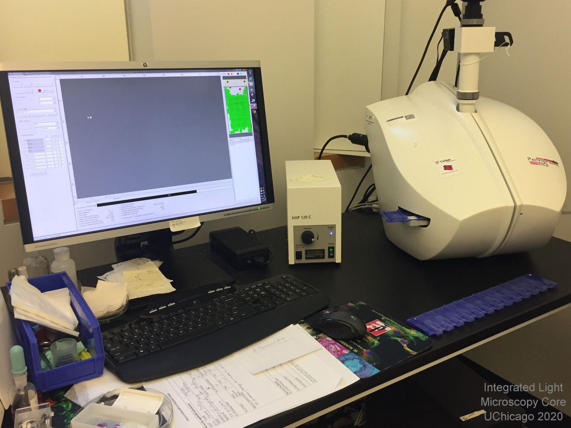 Overview: Slide scanning on the Pannoramic scanners is run as a drop off service. Our whole slide scanners from Cambrige Research and Instrumentation (CRi, now owned but NOT SUPPORTED by a spinoff of ThermoFisher) create a virtual library of histology or multi-channel fluorescent slides. Using the proprietary software available in the facility or as a free download, details on the virtual slide can then be zoomed to a virtual magnifications from 0.5x-100x and exported as .tif or .jpeg for analysis or presentation with other software packages.
Location: KCBD 1250E
Drop off Contact: Khalil Rodriguez
Fluorophores this microscope can image:
- Blue (e.g. DAPI)
- Green (e.g. GFP, Alexa 488)
- Red (e.g. mCherry, Alexa 594)
- Far Red (e.g. Alexa 647)
- Full color histology imaging with white light
Excitation Light Sources:
- HXP 120 C arclamp with fiber optic lightguide for fluorescence
- Incandescent white light bulb for histology
Emission Detection:
- Zeiss AxioCam MRm high sensitivity, 12-bit greyscale, 6.45um per pixel camera for fluorescence imaging
- CIS 3CCD 3 megapixel color camera for histology
Objective: 20x Zeiss LWD objective, virtual magnification range from 0.5x to 100x
Pixel Sizes: 0.3225 um/pixel (fluorescence) and 0.3225 um/pixel (histology)
Special Features:
- Drop off service: drop off your slides and put your name in the queue. We'll do the rest and notify you when your scans are done. Slides are scannned on a first-come, first-served basis.
- Automated scanning of up to 12 slides per batch
- Data is available on our server, so you can download it straight to any campus computer.
- Offline image viewing software available free from 3DHistech (Windows only) or analysis software free from QuPath at the University of Edinburgh (multi-platform: Windows, MacOS, Linux).