Stem cells (green) and their progeny (red) in the mouse colon
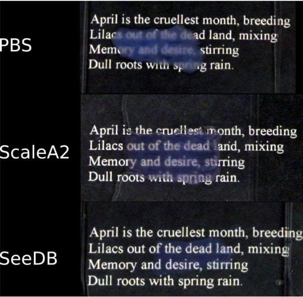
Turning tissue transparent for high-resolution imaging
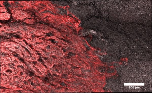
Imaging the interface of wound healing (red) next to a colonic ulcer
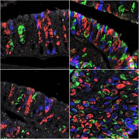
Tracking the fates of individual intestinal cells using multicolor reporter mice
Crypt “families” emerging during intestinal recovery from severe inflammation

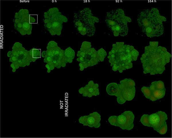
Laser-guided destruction of colonic “mini-guts” grown in a dish
Support cells (red) of the intestine in a genetically-modified mouse model

Neurons (red) highlighted during a video fly-through in the colonic wall
Click here to download video