Apr 13, 2020 | Immunology, Microbiome
by Helen Robertson
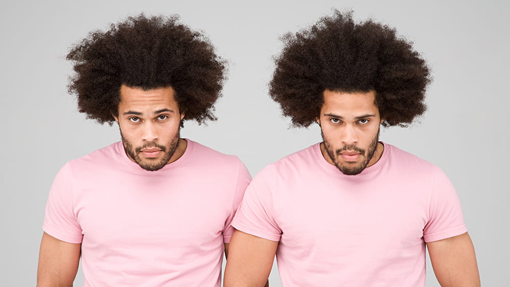
In 1933, identical twin baby boys Oskar Stohr and Jack Yufe were separated as a consequence of their parents’ divorce. Their subsequent upbringings could not have been more different: Oskar was brought up as a Catholic in Germany and became an enthusiastic member of the Hitler Youth. Jack remained in the Caribbean where they were born, was Jewish, and even lived for a time in Israel. Yet when they were reunited some fifty years after they last saw each other they had an uncanny number of similarities. They shared thought patterns, walking gait, a taste for spicy food, and perhaps most unusually, a habit of flushing the toilet before using it.
The unfortunate separation of identical twins provides scientists with an exceptional laboratory for exploring the “nature vs nurture” conundrum. How much of our identity is conferred by our genes, and how much is a product of the environment in which we are raised? This is also true for our microbiome: twin studies have shown that the microbiomes of identical siblings are far more similar than those of fraternal twins, indicating that genes are at play.
UChicago researchers Alexander Chervonsky, PhD, and Tatyana Golovkina, PhD, are particularly interested in exploring how genetics—especially the genes that control our immune system— influence the composition of our microbiome. They chose to explore this question with mice.
Both mice and humans have two types of immunity: innate, “inborn” immunity, the first line of defense against pathogens, and adaptive immunity, in which immune cells are trained by the specific pathogens they encounter to fight off the same bad guys in the future.
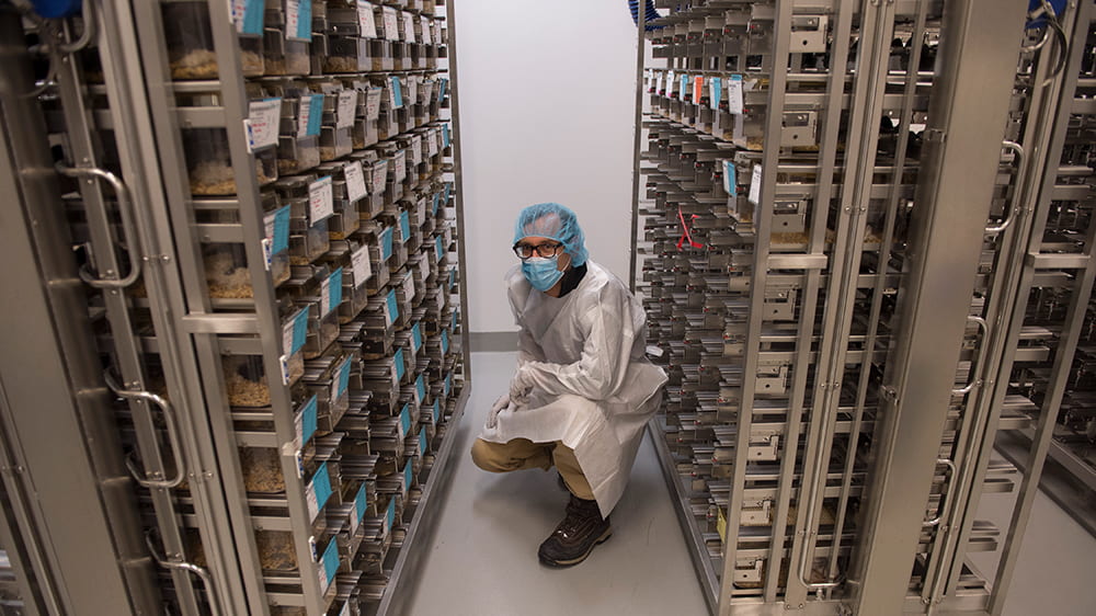
To make sure all the mice used in the study started with the same microbes, Chervonsky and Golovkina needed to isolate them from the regular, bacteria-filled world. A normal mouse—much like a normal human—is born into an environment with trillions of bacteria, spread to them from their mothers and cagemates, their handlers, bedding, and food. Fortunately, UChicago’s special germ-free “gnotobiotic” mouse facility allows scientists to experiment on mice born and raised in an environment that hosts precisely zero bacteria, which make the mice experimental blank slates.
The researchers transferred microbes from a source mouse, raised in a conventional environment, to several strains of germ-free mice: some genetically identical, others with slight differences in their immune-response genes. In collaboration with computational immunologist Aly A Khan, PhD, they compared the resulting microbiomes to see if changes to immune system genes resulted in different types of microbial communities.
They found that differences in the adaptive—targeted—immunity caused minimal differences in microbial composition, and that those differences affected only certain strains of bacteria. Other types of bacteria even took advantage of the genetic differences and multiplied.
The team was surprised to discover that it was the innate immune response—the one the mice were born with, that needs no training: it was more active in shaping the microbiome. But even then, the total influence over the microbes in the mice’s gut was fairly small, meaning there were likely other genetic and non-genetic factors at play in determining how bacteria colonized and proliferated in the animals guts.
The team intends to look deeper into understanding how genes and microbes influence each other in developing animals. But this study sets a valuable benchmark for future microbiome work: closely documenting how the immune system worked here in germ-free mice means comparisons against other studies are standardized. Now we have a better idea of the “nature” side of the equation.
The Gnotobiotic Research Animal Facility is a vital asset for researchers at UChicago, and just one example of the gold standard approaches being used by the Duchossois Family Institute to improve our understanding of the underlying components of health and wellness.
Helen Robertson is a postdoctoral scholar in Molecular Evolutionary Biology at the University of Chicago, with a keen interest in science communication and science in society.
Mar 11, 2020 | Microbiome
by Elise Wachspress
If you are a subscriber to the DFI blog (Easy! Free! See the box to the right!) or a regular reader, you’ve seen and heard a lot about mice. So much of what we have learned thus far about the interplay between the microbiome, immunity, and genetics comes from experiments with these little animals. Why?
Mice have historically been used in a lot of biomedical research. They are small and comparatively less costly to raise and house than other animals, reproduce quickly and often and have a short life span, thus studies can be conducted relatively quickly. Researchers have learned a lot about how to manipulate mice’s genetic inheritance, so the critters can make good models for some human diseases where we know the specific genes at work.
Mice have been especially useful for studying the effects of the gut microbiome, in diseases from obesity to cancer. Their intestinal track is similar to ours, with comparable anatomical features and many of the same kinds of cells—although there are some important differences.
Unlike humans, mice have a fore-stomach that holds food before digestion, creating an entry to the system much less acidic than our stomachs. Our intestines have more folds and a thicker mucus layer—an environment more friendly for bacteria looking to set up homes. The cecum, a part of the large intestine, is relatively larger in mice, offering a better place for microbial fermentation of indigestible fiber. And humans have an appendix, a place for bacteria to hang out when disaster (like diarrhea or other intestinal problems) strikes, the better to repopulate the intestines when the crisis is over.
Despite these caveats, mouse models have proven their value in gut microbiome studies, helping researchers understand some of the complex mechanisms that underlie diabetes, celiac, inflammatory bowel and others diseases. Mice offer huge practical advantages: researchers can readily control their diet, treat them with antibiotics, and transplant them with bacteria—unlike with humans, who are, none-the-less, the ultimate beneficiaries of these experiments.
Of course, transplanting mice with bacteria only works if the mice start out with either no bacteria or very well-characterized bacteria, so researchers can parse out the effects caused by the new microbial additions. After birth, the mice must continue to live their entire lives in an aseptic (no new bacteria) environment—and stay far away from others, as mice have a known habit of eating their cagemates’ poop. Managing this complicated endeavor is where Betty Theriault, DVM, and UChicago’s Gnotobiotic Research Animal Facility (GRAF) come in.
The word gnotobiotic is a mashup of two Greek roots, “known” and “alive.” The premise for the gnotobiotic facility is to understand exactly what microorganisms are alive in each mouse. The GRAF specializes in managing animals free of all organisms (bacteria, viruses, fungi and parasites) and gnotobiotic animals—those who have been intentionally inoculated with a well-defined set of microbes.
Established in 2007, the GRAF is now one of the largest and most successful academic gnotobiotic mouse facilities in the world, with over 90 flexible isolators. Theriault and her crew of 13 dedicated, full-time personnel maintain hermetically sealed, individually ventilated cages on rack systems—think sterile condominium units for mice—which assure each mouse is living with only the microbes it came in with. In 2017, the GRAF was named international laboratory animal team of the year award; you can take a three-minute tour of their approach on YouTube.
Theriault is a veterinarian with three decades of experience working with a broad spectrum of animal species in a variety of settings. After vet school at the University of California, Davis and an internship in Small Animal Medicine and Surgery at Penn, she came to UChicago to advance transplantation immunology research. Although she left for a while to pursue private practice, she returned to UChicago fifteen years ago to develop the GRAF, which is accredited by the Association for Assessment and Accreditation of Laboratory Animal Care International, a private nonprofit that promotes the humane treatment of animals in science. (You can watch Theriault present on the challenges involved in setting up a gnotobiotic facility at the National Academies of Sciences, Engineering, and Medicine in 2017).
So what’s the future of gnotobiotic research? Studies have shown that germ-free mice successfully support complex fecal microbiota from humans in their gastrointestinal tracks. Researchers anticipate mouse models will be increasingly useful in predicting patient outcomes to therapeutic interventions or informing personalized approaches to patient care.
While knowledge gained from animal models is not always directly transferable to humans, UChicago’s Gnotobiotic Research Animal Facility is helping us understand many more of the mechanisms at work between our gut microbes and our health, providing insights that may help us live longer, healthier lives.
Elise Wachspress is a senior communications strategist for the University of Chicago Medicine & Biological Sciences Development office

Feb 14, 2019 | Microbiome
by Folabomi Oladosu, PhD
Post-doctoral researcher specializing in pain and women’s health at NorthShore University HealthSystem
At the start of your doctor’s visit, as the nurse checks your pulse and blood pressure, you probably take slow deep breaths, trying to convince your doctor and yourself that you’re cool, calm, and collected. (No? Is that just me?)
Your blood pressure can tell your doctor a lot about you—including your risk for peripheral arterial disease, or PAD. In PAD, arteries that carry blood throughout the body are narrowed by deposits of fat and cholesterol. The disease is common, with more than three million new cases surfacing every year. PAD puts people at increased risk of both heart attack and stroke, so it’s important to treat it quickly and effectively.
When diet, physical activity, or medications are not enough, surgery is the next step to open up the narrowed arteries. Options include an angioplasty, where a tiny balloon is inserted into the blocked artery. The balloon is inflated to crush the plaque deposits and then removed to restore blood flow.
Following surgery, these once clogged arteries should carry red blood cells throughout the body like an amusement park water slide. To your dismay, you find this water park attraction might need more repairs three to 12 months later because of restenosis, the abnormal re-narrowing of arteries following surgery (Get it? Re-stenosis). Restenosis occurs in about 40 percent of patients after angioplasty and is usually treated with yet another surgery.
What causes this re-narrowing? Inserting the balloon and compressing the plaque may cause a mild arterial injury. The immune system responds, sending in white blood cells and platelets to repair the artery. But sometimes the immune system goes overboard and causes scarring instead. This means that the same arteries that were first narrowed by fatty deposits are narrowed yet again because of scarring caused by the immune system.
Why does the immune system to go into hyper-drive following surgery? One contributing factor may be—amazingly—the bacteria living in our gut. Drs. Eugene Chang and Betty Theriault of the University of Chicago collaborated with Dr. Karen Ho of Northwestern University to investigate the role of the gut microbiome on arterial healing following surgery.
Using their extensive knowledge and technical expertise, the team developed a method to study arterial healing in germ-free mice, mice with no gut bacteria. Their work revealed that germ-free mice developed much less arterial scarring following surgery compared to normal mice. Their research also showed that the immune system of germ-free mice was different than normal mice, using different white blood cells less likely to encourage swelling and scarring at the injury site.
All of us have been surrounded by germs since the day we were born. So if we need an angioplasty, we’d be like those normal mice, more likely to have arterial scarring after surgery.
Is there anything we can do to prevent restenosis? Related work by Drs. Ho and Chang suggests yes. A month-long treatment of the antibiotic vancomycin paired with sodium butyrate (a compound that slows cell growth) reduced arterial scarring in rats following an angioplasty. Legumes like beans and peanuts, when digested by gut bacteria, are a common source of sodium butyrate. This suggests that the dietary changes prior to angioplasty may help reduce arterial scarring and restenosis.
This “basic science” research by Drs. Chang, Theriault, and Ho provides great insights for better health. It shows that, despite their microscopic size, gut bacteria can greatly influence how we heal from common surgeries. The humble legume–cheap, tasty, easily stored, and environmentally friendly to grow—may help to keep our internal water slides open for service long after initial repairs.


