Areas of Research
The Internal Environment
(Individual neural, cognitive, and affective mechanisms)
Our ongoing work examining the internal environment makes use of neuroimaging methods, including fNIRS and fMRI, to study the brain. Read descriptions of some of these projects below.
The Neurological Underpinnings of Learning Math via Gesture-Based Instruction
Gesture–based instruction in math helps children retain and transfer what they learn. Most research investigating gesture is behavioral and does not address underlying mechanisms associated with its benefits. Some theorize that gesture is beneficial due to the incorporation of movement in the learning process; however, action–based instruction, which also involves movement, does not show these benefits. Our team is using functional near-infrared spectroscopy (fNIRS) to collect brain activty data while children receive either gesture– (movement with the hands) or action– (movement of objects in space) based instruction on math equivalence problems. We predict that gesture–based instruction will activate brain areas associated with relational reasoning and will be positively associated with solving problems requiring retention and transfer.
To read our past submission to sfNIRS, click here
To read about gesture-based instruction, click here
To read about fNIRS, click here


Work with the Adolescent Brain and Cognitive Development (ABCD) Study
The ABCD study is an ongoing study that follows the brain development of 10,000 nine- and ten-year-olds over ten years, collecting structural and functional MRI as well as behavioral data. This dataset enables groundbreaking human neuroscience research at unprecedented scale. The ENL has privileged access to these data, and a number of our recent projects have used the ABCD dataset to better understand how the environment affects brain development. Our relationship to these data is ongoing, and future lab members will have the opportunity to use the ABCD dataset for their projects.
To read about the ABCD study, click here
To read about ENL projects that have used the ABCD dataset, click here
To read about some of our previous work on the internal environment, see below.
Improvements in task performance after practice are associated with scale-free dynamics of brain activity
Although practicing a task generally benefits later performance on that same task, there are individual differences in practice effects. One avenue to model such differences comes from research showing that brain networks extract functional advantages from operating in the vicinity of criticality, a state in which brain network activity is more scale-free. We hypothesized that higher scale-free signal from fMRI data, measured with the Hurst exponent (H), indicates closer proximity to critical states. We tested whether individuals with higher H during repeated task performance would show greater practice effects. In Study 1, participants performed a dual-n-back task (DNB) twice during MRI (n = 56). In Study 2, we used two runs of n-back task (NBK) data from the Human Connectome Project sample (n = 599). In Study 3, participants performed a word completion task (CAST) across six runs (n = 44). In all three studies, multivariate analysis was used to test whether higher H was related to greater practice-related performance improvement. Supporting our hypothesis, we found patterns of higher H that reliably correlated with greater performance improvement across participants in all three studies. However, the predictive brain regions were distinct, suggesting that the specific spatial H↑ patterns are not task-general.


The role of preference in the emotional benefits of nature exposure
Functional near infrared spectroscopy (fNIRS) is a neuroimaging technique that measures the same biological signal as fMRI and can be used in a variety of less restrictive settings than an MRI scanner. In this study, participants completed an N-back working memory task while fNIRS data were recorded. As expected, participants showed greater fronto-parietal cortical activity during a more difficult 2-back task vs. an easier 1-back task. However, the results for the 3-back task were less consistent and it was hypothesized that this was due to somewhat low accuracy on the 3-back task. To see how task accuracy might be influencing the results, a behavioral partial least squares (PLS) analysis was conducted, which is a data-driven multivariate technique that has been used in other neuroimaging modalities to identify brain-behavior associations. Results of this analysis showed that the relationship between fNIRS activity (defined by change in deoxyhemoglobin concentrations (HbR) in mid-frontal cortex) and task accuracy does indeed differ depending on task difficulty (N-back level). More specifically, a larger reduction in HbR (equivalent to increased neural activity) was positively correlated with higher performance on the 3-back task, unrelated to activity on the 2-back task, and negatively correlated with performance on the 1-back task.
Evaluating Functional Localizers: The Case of the FFA.
Functional localizers are routinely used in neuroimaging studies to test hypotheses about the function of specific brain areas. The specific tasks and stimuli used to localize particular regions vary widely from study to study even when the same cortical region is targeted. Thus, it is important to ask whether task and stimulus changes lead to differences in localization or whether localization procedures are largely immune to differences in tasks and contrasting stimuli. We present two experiments and a literature review that explore whether face localizer tasks yield differential localization in the fusiform gyrus as a function of task and contrasting stimuli. We tested standard localization tasks–passive viewing, 1-back, and 2-back memory tests–and did not find differences in localization based on task. We did, however, find differences in the extent, strength and patterns/reliabilities of the activation in the fusiform gyrus based on comparison stimuli (faces vs. houses compared to faces vs. scrambled stimuli).


Pattern Classification of fMRI data: Applications for analysis of spatially distributed cortical networks
The field of fMRI data analysis is rapidly growing in sophistication, particularly in the domain of multivariate pattern classification. However, the interaction between the properties of the analytical model and the parameters of the BOLD signal (e.g. signal magnitude, temporal variance and functional connectivity) is still an open problem. We addressed this problem by evaluating a set of pattern classification algorithms on simulated and experimental block-design fMRI data. The set of classifiers consisted of linear and quadratic discriminants, linear support vector machine, and linear and nonlinear Gaussian naive Bayes classifiers. For linear discriminant, we used two methods of regularization: principal component analysis, and ridge regularization. The classifiers were used (1) to classify the volumes according to the behavioral task that was performed by the subject, and (2) to construct spatial maps that indicated the relative contribution of each voxel to classification. Our evaluation metrics were: (1) accuracy of out-of-sample classification and (2) reproducibility of spatial maps. In simulated data sets, we performed an additional evaluation of spatial maps with ROC analysis. We varied the magnitude, temporal variance and connectivity of simulated fMRI signal and identified the optimal classifier for each simulated environment. Overall, the best performers were linear and quadratic discriminants (operating on principal components of the data matrix) and, in some rare situations, a nonlinear Gaussian naïve Bayes classifier. The results from the simulated data were supported by within-subject analysis of experimental fMRI data, collected in a study of aging. This is the first study that systematically characterizes interactions between analysis model and signal parameters (such as magnitude, variance and correlation) on the performance of pattern classifiers for fMRI.
The Functional Connectivity Landscape of the Human Brain
Functional brain networks emerge and dissipate over a primarily static anatomical foundation. The dynamic basis of these networks is inter-regional communication involving local and distal regions. It is assumed that inter-regional distances play a pivotal role in modulating network dynamics. Using three different neuroimaging modalities, 6 datasets were evaluated to determine whether experimental manipulations asymmetrically affect functional relationships based on the distance between brain regions in human participants. Contrary to previous assumptions, here we show that short- and long-range connections are equally likely to strengthen or weaken in response to task demands. Additionally, connections between homotopic areas are the most stable and less likely to change compared to any other type of connection. Our results point to a functional connectivity landscape characterized by fluid transitions between local specialization and global integration. This ability to mediate functional properties irrespective of spatial distance may engender a diverse repertoire of cognitive processes when faced with a dynamic environment.

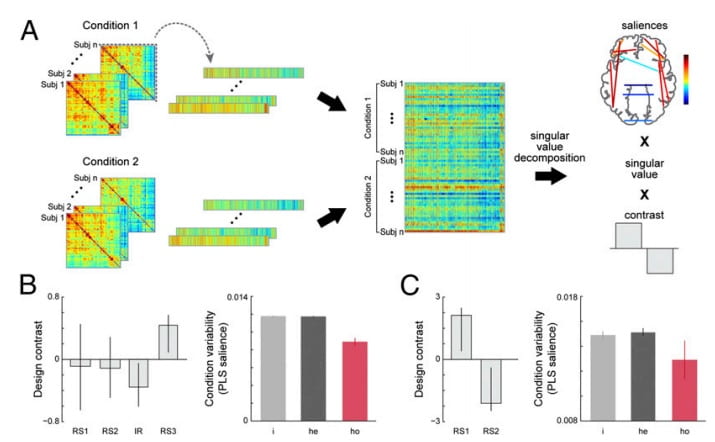
Stable long-range interhemispheric coordination is supported by direct anatomical projections
We show that the functional coordination between the two hemispheres of the brain is maintained by strong and stable interactions of a specific subset of connections between homotopic regions. Our data suggest that the stability of those functional interactions is mediated in part by the direct anatomical projections of large, highly myelinated fibers that traverse the corpus callosum. These functional properties were evident in both humans and macaques, suggesting a preserved framework for interhemispheric communication despite an increase in functional lateralization in humans. These results contribute to our fundamental understanding of how dynamic functional interactions between the two hemispheres of the mammalian brain are supported by its underlying anatomical architecture.
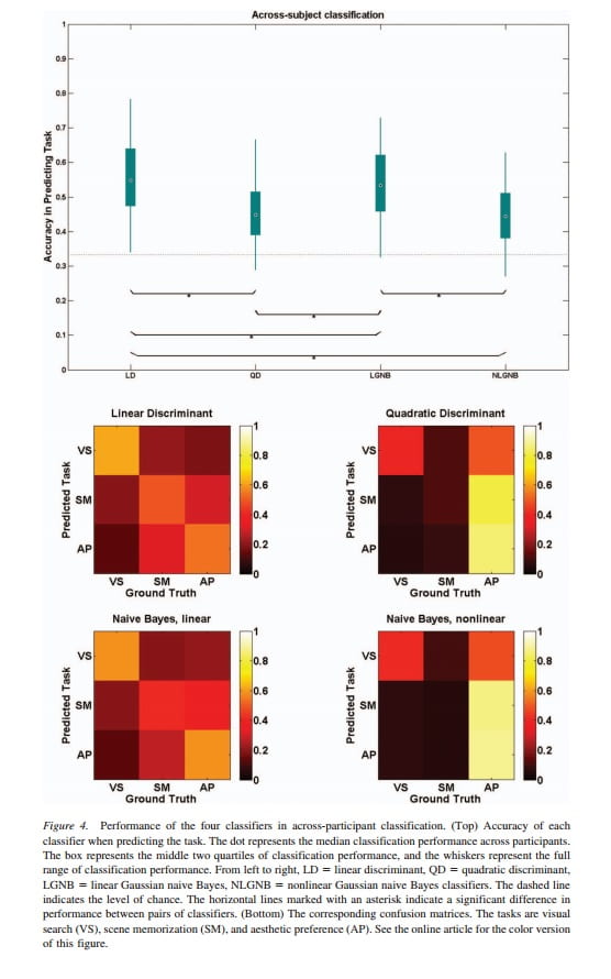
Classifying Mental States From Eye Movements During Scene Viewing
How eye movements reflect underlying cognitive processes during scene viewing has been a topic of considerable theoretical interest. In this study, we used eye-movement features and their distributions over time to successfully classify mental states as indexed by the behavioral task performed by participants. We recorded eye movements from 72 participants performing 3 scene-viewing tasks: visual search, scene memorization, and aesthetic preference. To classify these tasks, we used statistical features (mean, standard deviation, and skewness) of fixation durations and saccade amplitudes, as well as the total number of fixations. The same set of visual stimuli was used in all tasks to exclude the possibility that different salient scene features influenced eye movements across tasks. All of the tested classification algorithms were successful in predicting the task within a single participant. The linear discriminant algorithm was also successful in predicting the task for each participant when the training data came from other participants, suggesting some generalizability across participants. The number of fixations contributed most to task classification; however, the remaining features and, in particular, their covariance provided important task-specific information. These results provide evidence on how participants perform different visual tasks. In the visual search task, for example, participants exhibited more variance and skewness in fixation durations and saccade amplitudes, but also showed heightened correlation between fixation durations and the variance in fixation durations. In summary, these results point to the possibility that eye-movement features and their distributional properties can be used to classify mental states both within and across individuals.


Scale-Free Brain Dynamics Under Physical and Psychological Distress: Pre-treatment Effects in Women Diagnosed With Breast Cancer
Stressful life events are related to negative outcomes, including physical and psychological manifestations of distress, and behavioral deficits. Patients diagnosed with breast cancer report impaired attention and working memory prior to adjuvant therapy, which may be induced by distress. In this article, we examine whether brain dynamics show systematic changes due to the distress associated with cancer diagnosis. We hypothesized that impaired working memory is associated with suppression of “long-memory” neuronal dynamics; we tested this by measuring scale-free (“fractal”) brain dynamics, quantified by the Hurst exponent (H). Fractal scaling refers to signals that do not occur at a specific time-scale, possessing a spectral power curve P fð Þ/ f 2b; they are “long-memory” processes, with significant autocorrelations. In a BOLD functional magnetic resonance imaging study, we scanned three groups during a working memory task: women scheduled to receive chemotherapy or radiotherapy and aged-matched controls. Surprisingly, patients’ BOLD signal exhibited greater H with increasing intensity of anticipated treatment. However, an analysis of H and functional connectivity against self-reported measures of psychological distress (Worry, Anxiety, Depression) and physical distress (Fatigue, Sleep problems) revealed significant interactions. The modulation of (Worry, Anxiety) versus (Fatigue, Sleep Problems, Depression) showed the strongest effect, where higher worry and lower fatigue was related to reduced H in regions involved in visuospatial search, attention, and memory processing. This is also linked to decreased functional connectivity in these brain regions. Our results indicate that the distress associated with cancer diagnosis alters BOLD scaling, and H is a sensitive measure of the interaction between psychological versus physical distress.
Does resting-state connectivity reflect depressive rumination? A tale of two analyses.
Major Depressive Disorder (MDD) is characterized by rumination. Prior research suggests that resting-state brain activation reflects rumination when depressed individuals are not task engaged. However, no study has directly tested this. Here we investigated whether resting-state epochs differ from induced ruminative states for healthy and depressed individuals. Most previous research on resting-state networks comes from seed-based analyses with the posterior cingulate cortex (PCC). By contrast, we examined resting state connectivity by using the complete multivariate connectivity profile (i.e., connections across all brain nodes) and by comparing these results to seeded analyses. We find that unconstrained resting-state intervals differ from active rumination states in strength of connectivity and that overall connectivity was higher for healthy vs. depressed individuals. Relationships between connectivity and subjective mood (i.e., behavior) were strongly observed during induced rumination epochs. Furthermore, connectivity patterns that related to subjective mood were strikingly different for MDD and healthy control (HC) groups suggesting different mood regulation mechanisms.


Dimensionality of brain networks linked to life-long individual differences in self-control
The ability to delay gratification in childhood has been linked to positive outcomes in adolescence and adulthood. Here we examine a sub-sample of participants from a seminal longitudinal study of self-control throughout a subject’s life span. Self-control, first studied in children at age 4 years, is now re-examined 40 years later, on a task that required control over the contents of working memory. We examine whether patterns of brain activation on this task can reliably distinguish participants with consistently low and high self-control abilities (low versus high delayers). We find that low delayers recruit significantly higher dimensional neural networks when performing the task compared with high delayers. High delayers are also more homogeneous as a group in their neural patterns compared with low delayers. From these brain patterns, we can predict with 71% accuracy, whether a participant is a high or low delayer. The present results suggest that dimensionality of neural networks is a biological predictor of self-control abilities.
Neural and behavioral effects of interference resolution in depression and rumination
Individuals diagnosed with major depressive disorder (MDD) often ruminate about their depression and their life situations, impairing their concentration and performance on daily tasks. We examined whether rumination might be due to a deficit in the ability to expel negative information from short-term memory (STM), and fMRI was used to examine the neural structures involved in this ability. MDD and healthy control (HC) participants were tested using a directed-forgetting procedure in a short-term item recognition task. As predicted, MDD participants had more difficulty than did HCs in expelling negative, but not positive, words from STM. Overall, the neural networks involved in directed forgetting were similar for both groups, but the MDDs exhibited more spatial variability in activation in the left inferior frontal gyrus (a region critical for inhibiting irrelevant information), which may contribute to their relative inability to inhibit negative information.


Depression, rumination, and the default network
Major depressive disorder (MDD) has been characterized by excessive default-network activation and connectivity with the subgenual cingulate. These hyper-connectivities are often interpreted as reflecting rumination, where MDDs perseverate on negative, self-referential thoughts. However, the relationship between connectivity and rumination has not been established. Furthermore, previous research has not examined how connectivity with the subgenual cingulate differs when individuals are engaged in a task or not. The purpose of the present study was to examine connectivity of the default network specifically in the subgenual cingulate both on- and off-task, and to examine the relationship between connectivity and rumination. Analyses using a seed-based connectivity approach revealed that MDDs show more neural functional connectivity between the posterior-cingulate cortex and the subgenual-cingulate cortex than healthy individuals during rest periods, but not during task engagement. Importantly, these rest-period connectivities correlated with behavioral measures of rumination and brooding, but not reflection.