Register To Attend Before Time Runs Out!
Day(s)
:
Hour(s)
:
Minute(s)
:
Second(s)
What is the Rising Stars Symposium?
Diversifying the faculty in academic science takes more than just encouraging a diverse pool of post docs to apply to new faculty job postings. At its core a faculty cohort is a community, and communities grow and expand through relationship building. As the office of diversity and inclusion we are uniquely equipped to establish connections between scientists from diverse backgrounds with our own UChicago faculty. We hope that among these rising stars you will find, future collaborators, mentors and mentees, inspiration, and new ideas, not to mention potential faculty opportunities.
The Rising Stars Scholars are hand selected by department chairs. Each candidate’s research abstract is assessed for impact, innovation, and departmental alignment. The speakers chosen this year have emerged as the most interesting potential future faculty from a competitive field of abstract submissions. You won’t want to miss this.
The two-day virtual symposium culminates in a networking session from 3pm-5pm over zoom. Registrants may also request to be put in contact with speakers directly through the office of diversity and inclusion.
Register to attend here: https://redcap.link/103lf8mz
Meet the Rising Stars Scholars Below!


Ronald Fowle-Grider, PhD
Washington University
Talk title: Fructose Promotes Tumor Growth by Metabolite Exchange
Key words :Cancer metabolism, fructose, lysophosphatidylcholines, ketohexokinase, isotope tracing.
Research Abstract: Sugar and fructose consumption has increased considerably over the past five decades, largely due to the widespread use of high-fructose corn syrup (HFCS) as a sweetener. It has been proposed that fructose promotes the growth of some tumors by serving as a direct fuel source for cancer cells. Here, we show that diets enriched with HFCS or fructose water enhance tumor growth in animal models of melanoma, breast cancer, colon cancer, and cervical cancer without causing weight gain or insulin resistance. Interestingly, however, the cancer cells themselves were unable to use fructose readily as a nutrient because they did not express ketohexokinase-C (KHK-C). Primary hepatocytes did express KHK-C, which enabled fructose to be transformed into a variety of lipid species, including lysophosphatidylcholines (LPCs). The LPCs were excreted by hepatocytes and consumed by co-cultured cancer cells, which subsequently increased proliferation rates. Cancer cells used LPCs to generate a large proportion of their phosphatidylcholines (PCs), the major phospholipid of cell membranes. In vivo, tissues expressing KHK-C similarly transformed fructose into LPCs that were released into the circulation. With HFCS administration, serum levels of several LPC species increased by >7-fold. Administration of LPCs to mice was sufficient to increase tumor growth, and LPCs were depleted in tumor interstitial fluid. Even though PF-06835919, a drug previously in clinical trials to inhibit KHK-C in non-alcoholic fatty liver disease, had no direct effect on cancer cells, it decreased circulating LPC levels and prevented fructose-mediated tumor growth in vivo. Taken together, these findings reveal LPCs as a major metabolic destination of fructose metabolism and that an increase in fructose-derived nutrients such as LPCs can enhance tumor growth through a tumor cell non-autonomous mechanism.

Smriti Sangwan, PhD
University of California, San Francisco
Talk title: Co-translational mRNA surveillance by the ER stress sensor, IRE1
Key words: IRE1, ER protein homeostasis, cryoEM,
Research Abstract: IRE1, a membrane protein in the endoplasmic reticulum (ER) monitors the protein folding status of the ER and corrects the buildup of misfolded proteins. This process termed the unfolded protein response is responsible for the maintenance of protein homeostasis in all eukaryotic cells. IRE1 issues corrective action by two main pathways; by upregulation of chaperones through cleavage and expression of XBP1, a transcription factor and by reducing the burden of ER-targeted proteins by cleaving mRNAs. How IRE1 senses mRNAs in proximity for cleavage is currently unknown. Recent work from our lab suggests that IRE1 interacts with the co-translational machinery directly possibly sensing mRNAs on the ribosome but molecular details of this interaction are lacking. Here we present the composition of ribosomes that engage with IRE1 in mammalian cells using single-particle cryoEM. Under steady conditions, IRE1 interacts with translating ribosomes surveilling the mRNA for the presence of a consensus endomotif. However, under ER stress IRE1 engages with predominantly stalled ribosomes. IRE1 binds at the mRNA entry channel engaged with ribosomal proteins. Finally, through next- generation sequencing, we identified a core set of ER-targeted mRNAs that are recognized by IRE1. Our results reveal the mechanistic details of IRE1 interacting with the co-translational machinery.

Dominique Stephens, PhD
Fisk and Vanderbilt University
Talk Title: Elucidating Novel Pathways of Cancer Progression and Autoinflammation Immune Responses
Key words: Microscopy, Immune Response, Cancer Biology, Spatiotemporal Organization
Research Abstract: Inflammation in diseases characterized by dysregulated antiviral responses, is often triggered due to interactions between pro-inflammatory signals from the host or virus, known as DAMPs/PAMPs, and the body’s innate immune system. Host or viral nucleic acids, especially RNAs, can trigger the pro-inflammatory signaling cascade of the Retinoic acid-Inducible Gene-I (RIG-I). RIG-I is a pattern recognition receptor that identifies 5’ triphosphorylated RNAs and initiates the production of interferons (IFN), a class of proinflammatory cytokines. Modulating RNAs to be less detectable by RIG-I is an evolutionary strategy to prevent host autoimmunity, but it can unfortunately be exploited by pathogens to evade clearance. In this context, the RNA triphosphatase Dual Specificity Phosphatase 11 (DUSP11) plays a significant role. DUSP11 converts the 5’ triphosphate of RNA into a monophosphate, thereby modulating the sensitivity of autoinflammatory and antiviral cytokine responses. This suggests that DUSP11 may be instrumental in regulating the balance between immune response and pathogen evasion. Further, DUSP11 mediated modulation of the immune response may be applicable in other disease mechanisms, such as those found in the tumor microenvironment (TME).
In the context of cancer, it’s well-established that tumor cells manipulate immunomodulatory mechanisms to evade the immune response during tumorigenesis. It’s also recognized that viral infections can increase cancer risk and diagnosis due to the impact of viral RNA on host cell genes. This understanding has spurred research into the role of altered coding and non-coding RNAs in tumor growth. However, the potential role DUSP11 in tumorigenesis remains largely uncharted territory. While DUSP11 is known to modulate RIG-I response during viral infections, the mechanisms by which DUSP11 controls the inflammatory response in a viral or tumor context are not well understood. This highlights the need for further investigation into DUSP11’s role in regulating anticancer defenses during tumorigenesis.
My long-term goal is to understand how immunostimulatory biomolecules are controlled and use this knowledge to develop new treatments that can help regulate viral, inflammatory, and cancer-related conditions. This proposal will identify the mechanisms that control the immune modulation potential of DUSP11’s effect on the TME. My preliminary data demonstrate a significant redistribution of DUSP11 within cells in a co-cultured tumor microenvironment (involving fibroblast and tumor cell lines). This observation implies that DUSP11 may play a crucial role in the cellular response to the tumor environment, underscoring the importance of further exploration of its function in this context. My hypothesis is that DUSP11 regulates the sensitivity of inducing an inflammatory response by controlling RIG-I detection of immune-activating molecules.
Maria Fernanda Forni, PhD
Yale University
Talk title: Metabolic Regulation of Cell Fate and Function in Mammalian Skin: Implications for Tissue Homeostasis and Aging
Keywords: stem cells, immune cells, metabolism, regeneration, aging, wound healing, inflammation
Research Abstract :
Dr. Maria Fernanda Forni is a cell and molecular biologist and a Pew Postdoctoral Fellow at the Molecular, Cellular, and Developmental Biology Department in the Horsley Lab at Yale University. Her overarching goal is to understand how metabolism regulates cell fate and function. For that, she uses the skin as a model to investigate how stem and immune cells are regulated by energetic substrate availability and how metabolic changes regulate cell fate. She earned her BSc in Biology from the University of São Paulo and her Ph.D. in Biochemistry and Molecular Biology from the University of São Paulo with a period at the Stem Cell Center of Cambridge University, UK. As an independent scientist, I will focus on understanding the molecular mechanisms involved in the metabolic control of the immune and stem cell fate and function and how they collectively coordinate tissue homeostasis in mammalian skin. Her research program is focused on characterizing the unique metabolic landscapes displayed by stem and immune cells and how those change upon commitment to differentiation.
Another interest is to unveil how environmental shifts in the availability of energetic substrates impact the self-renew, homeostasis, and long-term maintenance of these cells. A core motivation driving this work is the search for metabolic signatures associated with normal homeostasis and how these become derailed during aging and disease. This can lead to future therapies to tackle aging itself and age-associated diseases such as overt inflammation and cancer. In pursuit of this goal, her research focuses on three complementary themes which aim to: (1) characterize the metabolic landscape and its contribution to the developmental morphogenesis of skin, (2) understand the metabolic profiles displayed during normal homeostasis and regeneration, and (3) what is the impact of metabolic changes during disrupted homeostasis such as aging, inflammation, and cancer. In summary, my research program will unveil very basic aspects of the metabolic control of mammalian cell fate and function and will pave the way for developing strategies to promote healthy aging and prevent age-related diseases.

Olusiji Akinrinmade, PhD
Albert Einstein College of Medicine
Talk title: Diapause-like adaptation with suppressed Myc activity enables tumor treatment persistence
Key words: Myc; adaptation to stress; drug persistence; residual tumor, diapause.
Research Abstract: Cytotoxic drugs often fail to completely eradicate tumors because of the viable cancer cells that persist during treatment (“residual tumors”) and represent a reservoir for tumor repopulation. The tumor cells that persist after therapy can remain dormant for months or years, until the eventual cancer relapse. The presence of residual cancer cells is strongly associated with aggressive disease and shortened progression free survival. Eliminating these persistent cells could significantly improve patient outcomes or even lead to cures; however, their biology is poorly understood and specific therapeutic agents targeting this clinical stage of the disease are not available. Our lab has generated in-vitro and in-vivo models of treatment persistence that faithfully recapitulate the phenotype and molecular profile of residual cancer cells, providing a robust preclinical tool to dissect their dependencies. We have shown that treatment-persistent cells in organoids, xenografts and in cancer patient biopsies adopt a distinct and reversible molecular program with suppressed Myc activity that resembles the dormant stage of embryonic diapause. This treatment-induced diapause-like cancer cell state is characterized by overall suppression of biosynthetic activity, reduced redox stress and attenuated apoptotic priming, which contribute to cancer cell survival during therapy. Our molecular and functional analyses strongly suggest that these chemo-persistent dormant tumor cells possess distinct genomic and pharmacological vulnerabilities that are not reflected by historical cancer models. We are currently using genomic (e.g. CRISPR-based approaches) and chemical (e.g. compound libraries) tools to identify the distinct dependencies of diapause-like cancer cells, which will allow the design novel therapeutic approaches to eliminate them. Targeting the mechanisms by which tumor cells enter, survive during, and exit this dormant diapause-like stage, opens new possibilities for the clinical management of cancer and increases the likelihood for cures.

Diego Pedroza, PhD
Baylor College of Medicine
Talk title: CSF-1R antibody targeting therapy with combined metronomic chemotherapy and immune checkpoint blockade enhance a B and T cell response to attenuate metastatic triple negative breast cancer.
Key words: Tumor associated macrophages, metastatic triple negative breast cancer, chemotherapy, immunotherapy, spatial biology
Research Abstract: Background: Increased macrophage infiltration and elevated levels of Epithelial to Mesenchymal Transition (EMT) gene signatures are associated with a poor outcome following neo-adjuvant chemotherapy in patients with triple negative breast cancer (TNBC). Further, metastatic disease with overall the poorest survival rates has a cold tumor immune microenvironment (TIME), especially in lung and liver metastases. Interestingly, an abundance of macrophages are observed at these metastatic sites. We have extensively characterized multiple preclinical syngeneic Trp53-/- tumor models that have extensive Tumor Associated Macrophage (TAM) infiltration. These tumor models transcriptionally resemble “claudin-low”, “basal-like” and “luminal-like” breast cancers. From these models the “claudin-low” closely phenocopy the high EMT/TAM subtype observed in patients. Objective: Accordingly, we asked if SNDX-ms6352 a novel specific, high affinity monoclonal antibody targeting CSF-1R in combination with Chemotherapy (CTX) and immune checkpoint blockade might provide an alternative approach for treating established lung and liver metastases. Methods: Single cells from primary T12 mammary tumors were introduced either via tail vein injection to obtain lung metastases or portal vein (PV) injection for liver metastases. Mice were injected with BrdU and multiple lung and liver tissues are harvested randomly to identify established metastases. The mice were randomized into treatment groups and administered four weekly treatments of IgG, CTX, SNDX-ms6352, CTX+SNDX-ms6352 or CTX+SNDX-ms6352+aPD1. Tissues were harvested and fixed overnight in 4% PFA then placed in 70% EtOH for paraffin embedding and stained for H&E, Immunohistochemistry (IHC), Immunofluorescence (IF) and Imaging Mass Cytometry (IMC). Results: Following four weekly treatments of IgG, SNDX-ms6352 and CTX+/-SNDX-ms6352 we observed a decrease in lung metastatic burden and an increase in overall survival as compared to IgG, CTX and SNDX-ms6352 alone. Interestingly we also observed increased CD8 T-cell infiltration, but only in the combination treated mice 28 and 56 days post treatment. However, lung metastases recurred in all of the mice several weeks later. Data from IMC showed intra-tumoral heterogeneity with increased PD-L1 and Ki67. Short term treated lung and liver metastases with the addition of aPD1 demonstrated increased CD20+ B and CD8+ T cell expansion and cleared most of the lung and liver metastatic burden. These results suggest that targeting macrophages enhances the immunostimulatory effect of metronomic CTX with immune checkpoint blockade in treating not only primary tumors, but also established lung and liver metastases via the activation of the tumor immune microenvironment. Supported by R01CA148761-12 and American Cancer Society – AutoZone Breast Cancer Postdoctoral Fellowship, PF-22-163-01-MM grants
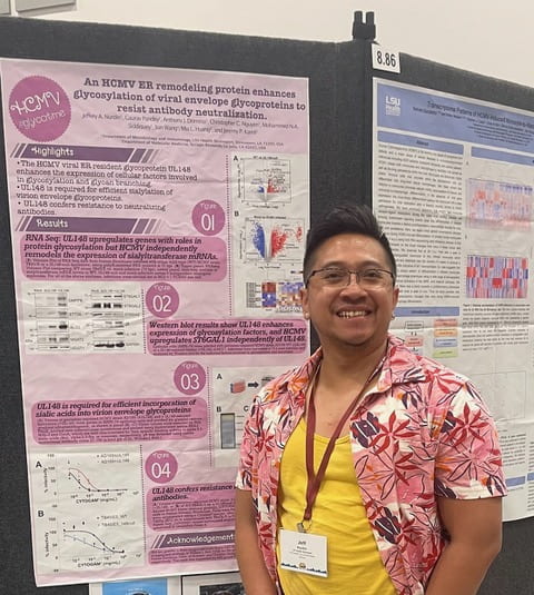
Jeffery Nurdin, PhD
Louisiana State University Health Shreveport
Talk title: Sugar? I barely even know her! HCMV glycoprotein upregulates host glycosylation to enhance neutralizing antibody resistance.
Keywords : Virology, immunology, glycosylation, humoral immunity, cytomegalovirus
Research Abstract:
Background: The glycosylation pathway contributes a diverse array of biological processes frequently exploited by enveloped viruses for glycoprotein folding, making glycan shields, or binding specific cell surface oligosaccharides for cell entry. However, whether viruses contribute to the glycosylation of their own glycoprotein is poorly understood. Human cytomegalovirus (HCMV) is a cosmopolitan dsDNA virus of the herpesvirus family that infects most human populations and persists for life. Despite asymptomatic presentation in most infected individuals, HCMV is a threat against immunocompromised patients and the leading cause of congenital birth defects. Therefore, elucidating mechanisms that contribute to efficacious vaccine development is urgently required. HCMV encodes a viral endoplasmic reticulum (ER)-resident glycoprotein UL148 that remodels the ER while activating an unfolded protein response (UPR). UL148 contributes to HCMV cell tropism by stabilizing viral envelope glycoprotein gO, which serves as the receptor binding subunit for gH/gL/gO that is essential for infectivity. This study found that UL148 also promotes glycosylation of envelope glycoproteins to resist antibody neutralization. Nonetheless, precisely how UL148 affects viral glycosylation is unknown and whether UL148 contributes to antibody evasion has not previously been explored.
Results: RNA sequencing (RNAseq) was performed to investigate the effect of UL148 on gene expression by infecting HFFT (human fibroblast cells) with HCMV strain TB40E in the presence or absence of UL148. RNAseq results demonstrate that UL148 is required during infection to significantly upregulate several cellular genes involved in protein glycosylation. Accordingly, Western blotting of HCMV virion strain AD169 repaired for UL131 and UL148 (AD169_r131+148) cultivated in ARPE-19 epithelial cells also exhibited decreased relative mobility of immunoreactive bands corresponding to gB, gH, gO, and UL130; compared to the virion from AD169_r131 without UL148, suggesting the difference of viral glycoprotein processing. AD169_r131 and AD169_r131+148 or TB40E and TB40E_148stop were incubated with serially diluted HCMV hyper-immune globulin (Cytogam) and overlaid onto HFFT or ARPE-19 cells to examine the effect of UL148 towards antibody neutralization. Moreover, virions produced in cells treated with UPR small molecule inhibitors showed antibody sensitization, suggesting that UL148-induced UPR activation plays a role in generating antibody-resistant virions. Collectively, UL148 influenced viral glycoprotein maturation resulting in immune-evasive virions. This discovery is proof that viruses indeed modulate the glycosylation of their own glycoproteins and could be harnessed to generate immune-sensitive virions for a better antibody response towards infection that elicits a better viral clearance.
Gina Gallego-Lopez, PhD
Morgridge Institute for Research-University of Wisconsin-Madison
Talk Title: Understanding The Host Metabolic Changes Induced by Apicomplexan Parasites Toward its Association to Cancer.
Research Abstract: Intracellular bacteria and protists are auxotrophic for many metabolites and must rely on the host cell to supply these nutrients. The mechanisms of how pathogens manipulate host metabolism to their benefit are not understood. These questions are difficult to address for intracellular pathogens because one cannot easily distinguish the origin of the metabolite as host or pathogen derived. Toxoplasma gondii, the causative agent of toxoplasmosis, is an obligate intracellular parasite that infects warm-blooded vertebrates across the world. In humans, seropositivity rates of T. gondii range from 10% to 90%. Despite its prevalence, few studies address how T. gondii infection changes the metabolism of host cells.
Toxoplasma gondii manipulates the host cell by a pre-invasion process called “kiss and spit”, where the contents of the parasite rhoptry organelles are secreted into the host cytoplasm before invasion occurs. This research project demonstrated that rhoptry contents from kiss and spit altered metabolite abundance in nucleotide synthesis, the pentose phosphate pathway, glycolysis, and amino acid synthesis. An increase in 2,3-bisphosphoglycerate (2,3-BPG) abundance led us to investigate the activation of host cytosolic nucleosidase II (cN-II) to provide purines for the parasite.
Additionally, Optical metabolic Imaging (OMI) was used to analyze the fluctuations in redox biology in alive infected cells. We investigate how T. gondii manipulates the host cell metabolic environment by monitoring metabolic response over time using non-invasive autofluorescence lifetime imaging of single cells, seahorse metabolic flux analysis, reactive oxygen species (ROS) production, and metabolomics. Autofluorescence lifetime imaging indicates that infected host cells become more oxidized and have an increased proportion of bound NAD(P)H with infection. These findings are consistent with changes in mitochondrial and glycolytic function, decrease of intracellular glucose, fluctuations in lactate and ROS production in infected cells over time. We also examined changes associated with the pre-invasion “kiss and spit” process using autofluorescence lifetime imaging, which similarly showed a more oxidized host cell with an increased proportion of bound NAD(P)H over 48 hours. Glucose metabolic flux analysis indicated that these changes are driven by NADH and NADP+ in T. gondii infection. In sum, metabolic changes in host cells with T. gondii infection were similar during full infection, and kiss and spit. Autofluorescence lifetime imaging can non-invasively monitor metabolic changes in host cells over a microbial infection time-course. Many of these host changes induced by this protozoa parasite are similar to cancer cells metabolism.

Valeria M Reyes Ruiz, PhD
Vanderbilt University Medical Center
Talk title: Exploiting Staphylococcus aureus signal transduction to identify novel antibacterial effectors of the macrophage
Key words: S. aureus, TCS, macrophage, CRISPR
Research Abstract: Staphylococcus aureus is a leading cause of morbidity and mortality. It is the leading cause of skin and soft tissue infection, endocarditis, and a frequent agent of bloodstream infections. While S. aureus is mainly considered an extracellular pathogen, recent studies describe an intracellular reservoir of S. aureus, which is poorly accessed by antibiotics. Moreover, S. aureus that grows and persists inside macrophages can disseminate and cause disease in mouse models of infection. Studies examining the interaction of intracellular S. aureus with macrophages would provide insight into the development of therapeutics to treat this bacterial reservoir, which circumvents antibiotic efficacy. S. aureus has evolved intricate regulatory networks that allow the bacterium to produce a diverse array of virulence factors and defense mechanisms within the host environment. These include two-component signal transduction systems (TCSs), in which a histidine kinase (HK) responds to extracellular stimuli and transfers a signal to a response regulator (RR) that mediates the regulation of gene expression. S. aureus contains 16 TCSs that respond to a diverse array of environmental signals. We leveraged the biology of TCS signaling to use S. aureus as a biosensor for the identification of novel antibacterial effectors of the macrophage.
We have engineered reporter constructs for each of the 16 TCSs in S. aureus. Through the application of advanced imaging modalities, we identified three S. aureus TCSs that are activated upon sensing host-imposed stresses inside macrophages. We then developed an arrayed genome-wide CRISPR screen in macrophages with the use of robotics and high-content imaging to discover host factors responsible for the control of the intracellular reservoir of S. aureus and for the activation of these regulatory systems. Our data analysis pipeline relies on the use of machine learning with robust segmentation to measure both TCS-dependent reporter signal and constitutive bacterial signal on a per-cell basis for every well in our arrayed library. This screen has revealed host gene products that affect TCS activation and bacterial burdens. Some of these host gene products are involved in the innate immune control of other infectious agents whereas others have no previously characterized function in antibacterial defense. My postdoctoral work is providing a foundation for a diverse and dynamic research program to determine the mechanism by which macrophage factors are mediating innate immune control of bacterial infection and activation of TCSs. These studies will elucidate novel innate immune factors required for the control of bacterial infections, as well as reveal the bacterial signaling pathways essential for overcoming host-imposed stress.

Y Hoang, PhD
University of Michigan
Key words: phase separation, positioning system, carboxysomes, microscopy, bacterial microcompartments
Research Abstract:
The carboxysome is responsible for sequestering and fixing almost 35% of the Earth’s atmospheric CO2. In cyanobacteria and chemoautotrophic proteobacteria, carboxysomes are equidistantly spaced along the cell length through the McdA and McdB proteins. Without this system, carboxysomes coalesce into a single aggregate, disrupting homeostasis and leading to cell death. Although functional carboxysomes have been expressed in heterologous hosts, their improper positioning leads to a significant decrease in carbon fixation rate due to the loss of substrate/product permeability and asymmetric carboxysome inheritance. Here, we reconstitute a minimal and self-organizing two-component system, the McdAB system, which is both necessary and sufficient to position carboxysomes in E. coli to maximize their inheritance and increase carbon fixation. We first confirmed the expected binding of McdA to the E. coli nucleoid by expressing a fluorescent fusion of McdA. Expression of McdB alone revealed its ability to undergo Liquid-Liquid-Phase-Separation, which we assessed using fluorescence microscopy and single particle tracking techniques. Intriguingly, coexpression of McdA and McdB resulted in the distribution of multiple McdB foci along the cell length, providing a foundation for engineering of the first self-organized droplet-positioning system in bacterial cells. Subsequently, by carefully modulating the levels of McdA and McdB during coexpression with the carboxysome operon, we successfully achieved equidistant positioning of previously polar aggregated carboxysomes onto the E. coli nucleoid. This was confirmed through fluorescence images, where Rubisco was labeled to visualize the carboxysome compartments, as well as Transmission electron microscopy (TEM) images. The successful reconstitution of the positioning system McdAB in E. coli establishes a platform for future positioning of other bacterial microcompartments in heterologous hosts, maximizing their inheritance and increasing yields of associated products.
Hananeh Fonoudi, PhD
Northwestern University, Feinberg School of Medicine
Talk title: Utilizing induced pluripotent stem cells as a tool to study complex human cardiovascular defects.
Key words : Cardiovascular defects, CRISPR/Cas9-based genome editing, High throughput drug screening, Cardiac organoids, Human induced pluripotent stem cells.
Research Abstract Withheld for Publication

Xiaotang Lu, PhD
Harvard University
Talk title: Bestowing Multiplexed Spatial Proteomic Imaging with Ultrastructural Context
Key words: Multiomics, Microscopy, Imaging
Research Abstract:
Heterogeneity characterizes many biological systems, from embryos and gut microbiomes to tumor microenvironments. As spatial omics rise as a pivotal tool, they reveal the intricate organization of diverse cell types within these systems. Yet, to truly grasp the unique working principles of these systems, a detailed description of their architecture is essential. In the nervous system, for example, distinct cell types with unique molecular characteristics form massively interconnected networks, collectively underpinning its vast functionality. My research, supported by the BRAIN Initiative K99, aims to integrate proteomics with ultrastructural imaging to understand the nervous system. I am also enthusiastic about extending this methodology to investigate other biological systems.
Postdoctoral Work:
Volumetric electron microscopic (EM) emerges as a comprehensive way of reconstructing neurons and mapping wiring diagrams, given its unique capability to unambiguously visualize synapses. Using correlated light and electron microscopy, we can overlay molecular information onto EM volumes, thus enhancing the interpretation of reconstructed neuronal circuits. Traditional immunofluorescence involves cell permeabilization, using detergent to dissolve membranes to allow antibodies to access intracellular epitopes. This process inevitably compromises tissue ultrastructure. My research has demonstrated that small engineered immunoprobes, including nanobodies from alpaca heavy-chain only antibodies1 and single-chain variable fragments of full-size antibodies2, can permeate cell membranes without using detergent. As a result, we can superimpose multiple fluorescent immunolabels onto ultrastructure-preserved EM images, revealing the molecular identities of brain cells, the types of synaptic connections, and the subcellular locations of ion channels.
With the surge in biomarkers discovered through single-cell sequencing, the pressing challenge is to expand the panel of labeling reagents. Traditional antibody screening is both labor- and time-intensive. My recent work highlighted single-stranded DNA-based aptamers as a solution3. Modified aptamers, like immunoglobulins, offer specific and high-affinity immunolabeling. Their small size facilitates detergent-free staining. Additionally, the ease of creating vast libraries of randomized DNA sequences for in vitro selection hints at the huge potential for multiplexed proteomic imaging.
Planned Future Research:
Efficiently expanding the aptamer library is essential. Often, antibodies selected for native proteins compromise their labeling efficiency due to antigen masking caused by fixation. Sourcing an antibody panel for immunohistochemistry remains challenging. My approach will leverage the aptamer platform, integrating tissues with varying fixations into the SELEX process. This approach promises compatibility for selected aptamers with PFA, FFPE, and hydrogel-embedded tissues (like CLARITY and Expansion Microscopy), enhancing the versatility of the aptamer imaging platform.
While typical immunofluorescence allows the detection of 4-5 protein targets, DNA-barcoded antibodies enable the assessment of many dozens of protein markers but require cumbersome antibody treatment and non-specific DNA conjugation. Aptamers present a streamlined barcode integration. This not only paves the way for advanced multiplexed imaging, including fluorescence imaging and in situ sequencing but importantly, this method aligns with EM, offering a holistic view of tissue architecture.
I am committed to building a versatile, multiplexed, and multimodal imaging platform that serves a broad spectrum of cell biology and clinical investigations, shedding light on the intricate molecule-structure interactions, their evolution over the lifespan, and disease-induced alterations.

Najla Arshad, PhD
Yale University School of Medicine
Ongoing research: I am currently investigating the dysregulation of antigen presentation by MHC-I by cancers and viruses. My research showed that SARS-CoV2-derived proteins downregulate MHC-I expression using distinct mechanisms. While ORF3a disrupted global protein trafficking in the secretory pathway, ORF7a specifically downregulated MHC-I. I discovered that ORF7a associates with the heavy chain and impacts MHC-I trafficking, acting as a molecular mimic of b2m. In the context of cancers, I study myeloproliferative neoplasms (MPNs), a type of chronic, incurable blood neoplasms. My model system is a subset of MPNs driven by frame-shift mutations in the chaperone protein calreticulin (CALR). CALR is primarily involved in glycoprotein folding in the endoplasmic reticulum but is also an essential part of a protein complex that loads peptides onto MHC-I. I showed that MPN-associated mutant CALR downregulated antigen presentation by MHC-I as a potential means of immune evasion. Ironically, this led to the presentation of tumor-associated peptides, a subset of which are immunogenic. I am following up on this translationally relevant finding to develop nanobody- and cell-based therapies to treat this subset of MPNs that have no targeted treatment.
Future research: Several chaperone proteins in the endoplasmic reticulum have been appropriated to aid in peptide loading and quality control of the MHC-I antigen presentation pathway. However, several other immunomodulatory proteins utilize these chaperones or the secretory pathway for their function. In the long term, I aim to study the impact of viral and cancer-induced perturbations of the ER and its chaperones on immune responses.

Vivian Lee-Kim, PhD
Brigham and Women's Hospital
Background: Coronary artery disease (CAD) is the leading cause of mortality world-wide, and is heritable. Genome-wide association studies (GWAS) identifies the 10q23 locus as a CAD risk locus that is independent of traditional CAD risk factors such hyperlipidemia, hypertension, and diabetes, and therefore provide an opportunity to discover novel molecular pathways and therapies for CAD. Here, we identified CAD risk variants in the 10q23 locus that affect tetraspanin14 (Tspan14) gene expression in endothelial cells (ECs) and demonstrate a role for TSPAN14 in EC function through the Notch signaling pathway.
Abstract Withheld for Publication
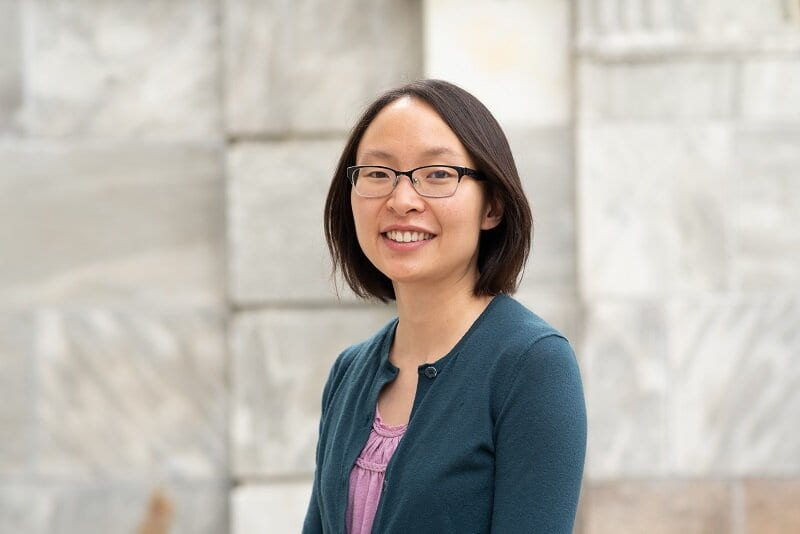
Janet Song, PhD
Boston Children's Hospital
Key words: human brain evolution, gene regulation, neural progenitors, human-chimpanzee tetraploids, CRISPRi screens
Research Abstract:
Although comparisons of human and non-human primate brains have identified thousands of molecular differences, it has been difficult to identify the human-specific sequence variants that underlie the dramatic modifications to brain size, connectivity, and function found in humans. One hurdle is that current comparative approaches cannot distinguish cis-regulated genes, which change in expression due to nearby sequence variants on the same DNA molecule, from trans-regulated genes, which change in expression due to changes in diffusible factors in the cellular environment (like the levels of cis-regulated transcription factors (TFs)).
To distinguish cis from trans changes, we generated human-chimpanzee tetraploid stem cell lines as a genetic model where the human and chimpanzee genomes are in the same cellular environment and only cis-regulated changes are observed. We have now used this system to profile cis– and trans-regulated genes (RNA-seq) and open chromatin peaks (ATAC-seq) by comparing human, chimpanzee, and human-chimpanzee neural progenitor cells (NPCs). Genes that are more highly expressed in humans are enriched for processes related to mitosis, consistent with increased neurogenesis in humans. Intriguingly, we identified cis-regulated TFs, including JDP2 and EBF1, whose motifs are enriched at trans-regulated open chromatin peaks, suggesting that these TFs may be major drivers of epigenomic and transcriptomic rewiring between human and chimpanzee NPCs.
To identify human-specific variants that underlie cis-regulated gene expression changes, we linked cis-regulated open chromatin peaks that contain derived sequence changes in humans to nearby cis-regulated genes. A CRISPR inhibition screen of 106 cis-regulated peaks identified species-specific enhancers, including ones near ANK3 and PROM1. Further characterization of cis-regulated TFs and non-coding regions, as well as the application of this model to additional cell types and paradigms, will advance our understanding of how human-specific sequence changes contribute to phenotypes that have arisen in the human lineage.

Fabian Morales-Polanco, PhD
Stanford University
Key words: Proteostasis, Misfolding, Aggregation, ESCRT, Nuclear-Vacuolar junctions
Research Abstract:
Misfolded proteins can acquire toxic conformations, disrupting essential cellular processes and leading to human diseases ranging from neurodegeneration to cancer. To prevent this, cellular proteostasis is maintained by a network of chaperones and clearance factors promoting protein quality control (PQC). One fundamental PQC strategy is to spatially sequester misfolded proteins into distinctly localized membrane-less compartments. PQC compartments segregate and concentrate damaging conformers enhancing their refolding or clearance through either the ubiquitin-proteasome system (UPS) or endo-lysosomal pathways. Misfolded cytoplasmic proteins partition into distinct inclusions depending on their aggregation state. Insoluble amyloid proteins are sequestered into the Insoluble Protein Deposit (herein IPOD), while soluble misfolded proteins (and not yet terminally aggregated) are sequestered into small, dynamic, ER-associated structures called Q-bodies which, during sustained stress, coalesce along the endomembrane system into a Juxtanuclear Quality Control compartment (herein the JUNQ). Spatial PQC of cytoplasmic proteins through the IPOD and JUNQ pathways is evolutionarily conserved from yeast to mammalian cells. In the nucleus, misfolded proteins are sequestered in a membrane-less intranuclear quality control compartment (herein INQ), proximal to the nucleolus.
We here examined the relationship between cytoplasmic and nuclear spatial PQC. Many studies showed cytoplasmic misfolded proteins being concentrated into the cytoplasmic JUNQ and cleared through an ER-anchored cytoplasmic UPS pathway. It has also been proposed that clearance of cytoplasmic misfolded proteins requires import into the nucleus, whereby the JUNQ would form an inclusion identical to the INQ. Addressing this issue, we unequivocally demonstrate that the JUNQ is cytoplasmic and the INQ is nuclear. Notably, a fascinating convergence phenomenon between these two compartments has been unveiled, as the INQ and JUNQ align themselves across the nuclear envelope, establishing a dynamic bridge for inter-compartmental communication. At the epicenter of this convergence lies the Nuclear-Vacuolar Junction (NVJ), a critical nexus connecting the nuclear envelope and the vacuole. Herein, the perinuclear ESCRT-II/III protein Chm7 plays a pivotal role, orchestrating the interaction of INQ and JUNQ with this strategic cellular junction. Moreover, we further demonstrate that the NVJ serves as a platform that facilitates VPS4-dependent vacuolar clearance, a fundamental mechanism for eliminating misfolded proteins. This clearance process involves an enthralling twist: nuclear misfolded proteins encapsulated within the INQ are extruded from the nucleus into the vacuole, highlighting the remarkable versatility of cellular architecture in adapting to the challenges posed by protein misfolding.
The notion that nuclear-vacuolar junctions act as hubs for spatial PQC reveals a novel facet of cellular architecture in proteostasis maintenance. (Figure 1). These insights not only enhance our understanding of fundamental cellular processes but also have broader implications for human health. Dysfunction in nuclear envelope dynamics and nucleocytoplasmic trafficking has been associated with various pathological conditions, including aging and neurodegenerative disorders. Our discoveries suggest a potential link between compromised nucleocytoplasmic trafficking and impaired misfolded protein clearance, contributing to the onset and progression of cellular dysfunction in these disorders.

Lisa Eshun-Wilson, PhD
The Scripps Research Institute
Talk title: Structural snapshots of AAA+ protease YME1 reveal substrate-free ADP-bound states that are proteolytically inactive
Key words: Protease; Enzymatic Activity; AAA+; ATPase; cryo-EM; conformational landscape.
Research Abstract Withheld for Publication

Cecilia Arriagada, PhD
Rutgers University
Talk title: Role of mesodermal Fibronectin in mechanotransduction during cardiac development
Key words: Cardiovascular development, mechanotransduction, ECM, Fibronectin
Research Abstract: Defective outflow tract (OFT) elonga&on causes congenital heart disease (CHD), resul&ng in newborn lethality. The OFT lengthens by the addi&on of progenitor cells derived from the second heart field (SHF). SHF cells in the splanchnic mesoderm are organized into an epithelial-like layer forming the dorsal pericardial wall (DPW). Tissue tension within the DPW, cell polarity, prolifera&on, and migra&on are requisite processes controlling the addi&on of SHF-derived cells to the heart and elonga&ng OFT. However, genes regula&ng these processes are not en&rely understood. Fibronec&n (Fn1) is an extracellular matrix glycoprotein essen&al for cardiac development, including the OFT. Although the requirement for Fn1 in OFT elonga&on is clear, the mechanisms underlying the role of Fn1 in OFT development remain unknown. We ablated Fn1 from the anterior mesoderm, including the SHF, using the Mesp1Cre/+ knock-in strain of mice. We show that Fn1 synthesized by the SHF is a central regulator of the epithelial architecture in the DPW. Fn1 expression is enriched in the anterior DPW and mediates ou*low tract elonga&on by regula&ng cell polarity, shape, cohesion, and mechanotransduc&on. Together, we establish that mesodermal Fn1 coordinates mul&ple cellular behaviors in the anterior DPW required for the elonga&on of the OFT and embryonic viability. This work leads to a deeper understanding of mechanisms regula&ng cardiac development and altera&ons that cause CHD.

Huan Meng, PhD
Baylor College of Medicine
Talk title: Temporal and Spatial Regulation of Metabolism and Stress Responses
Key words: 12-hour clock; NAFLD; prediabetic syndromes; NASH; cancer metabolism
Research Abstract: Dr. Meng’s research focuses on the mechanisms of metabolic regulation and clock control in metabolic physiology and their dysregulation in metabolic diseases and cancer metabolism. One major aspect of Dr. Meng’s research is the study of a newly discovered 12-hour circatidal clock and its contributions to the development and progression of liver metabolic diseases, diabetes, obesity, and cancer metabolism. His recent findings have revealed the existence of this clock, which operates independently of the 24-hour circadian clock. This 12-hour clock is cell autonomous and regulated by molecular pacemakers distinct from those controlling the 24-hour clock. He identified the transcriptional factor XBP1 and coregulator SRC-3 as key regulators of this 12-hour clock, which plays a crucial role in lipid metabolism. Both XBP1 and SRC-3 are oncogenes and metabolic drivers. His work has revealed the involvement of mammalian 12-hour clock regulators in modulating lipid membrane fluidity and mitochondrial functions. Notably, Dr. Meng has identified a novel role of the 12-hour clock in nonalcoholic fatty liver disease (NAFLD) and prediabetic syndromes. In addition, he has made significant advancements in understanding the spatial-temporal distribution of metabolic pathway enzymes in the nucleus and their biological function. Dr. Meng’s recent findings have established links between the temporal and spatial regulation of lipid metabolism and various metabolic diseases, as well as certain types of cancer that currently lack effective treatments. Using various advanced techniques such as single-cell multiomic RNA-Seq, ATAC-Seq, spatial transcriptomics, metabolomics, and epigenomic analyses, he investigates reprogramming mechanisms of signaling and nuclear factors on chromatin. By deciphering their role in reprogramming hepatic transcription and metabolism machinery, Dr. Meng aims to gain insights into the development of nonalcoholic steatohepatitis (NASH) and hepatocellular carcinoma (HCC). His multidisciplinary studies have important implications for understanding metabolic diseases, cancer metabolism, and aging. Ultimately, his work aims to target the crosstalk between the genome/epigenome, nutrition, and environment for the development of innovative therapeutics.

Kavita Rangan, PhD
UC San Diego
Talk title: Protein diversification and adaptation through RNA recoding
Key words: RNA editing, epigenetics, cephalopods, adaptation
Research Abstract:
I am interested in the epigenetic mechanisms organisms use to generate phenotypic plasticity and adapt in the face of changing environments. RNA editing of mRNAs occurs across all domains of life and is a remarkable example of how genetic information can be dynamically “recoded”; yet how this process supports organismal plasticity in response to environmental change is largely unexplored. My recently published work used microtubule motor proteins as a model to demonstrate that cephalopods use RNA editing to tune protein function in different tissues and in response to environmental temperature. My research vision builds off this work to ask broadly: how does RNA editing in diverse organisms contribute to phenotypic plasticity and adaptation? My research program seeks to 1) investigate how RNA recoding modifies protein function to support cellular processes across different environments 2) understand the molecular mechanisms regulating the extraordinary levels of RNA recoding in cephalopods and 3) explore how RNA editing contributes to genetic adaptation in filamentous fungi. Centered on RNA editing as a phenomenon, this research has the potential for diverse impact. Interrogating protein function through the lens of RNA recoding could yield novel therapeutic and biotechnology targets. Understanding the basic biology of non-mammalian RNA editing systems could impact the development of new site-directed RNA editing strategies. Climate change is a ubiquitous environmental stressor in the Anthropocene. Exploring how complex eukaryotes use RNA editing to thrive in diverse environments offers a window into how conserved cellular machineries can adapt and function across dynamic environments. Finally, understanding how fungi adapt to temperature and other environmental stress could help limit emerging plant and human pathogens.


