
Selective / Single Plane Illumination Microscopy (SPIM) aka Lightsheet Microscopy
 What is SPIM? Selective / Single Plane Illumination Microscopy is particularly suited to long-term timelapse imaging of larger living specimens, such as zebrafish, Drosophilia embryos, and small plants. This technology has also been used to image living and fixed 3D cell cultures, including organoids, cysts and 3D cell migration assays.
Hands-on Microscopes
To access a microscope, click the New User Training button above and work through our training checklist. Trained users have 24/7 access to the facility and are given permission to schedule their own microscope sessions though our online scheduling software.
LaVision Ultramicroscope II Large Format Upright Lightsheet
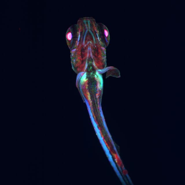
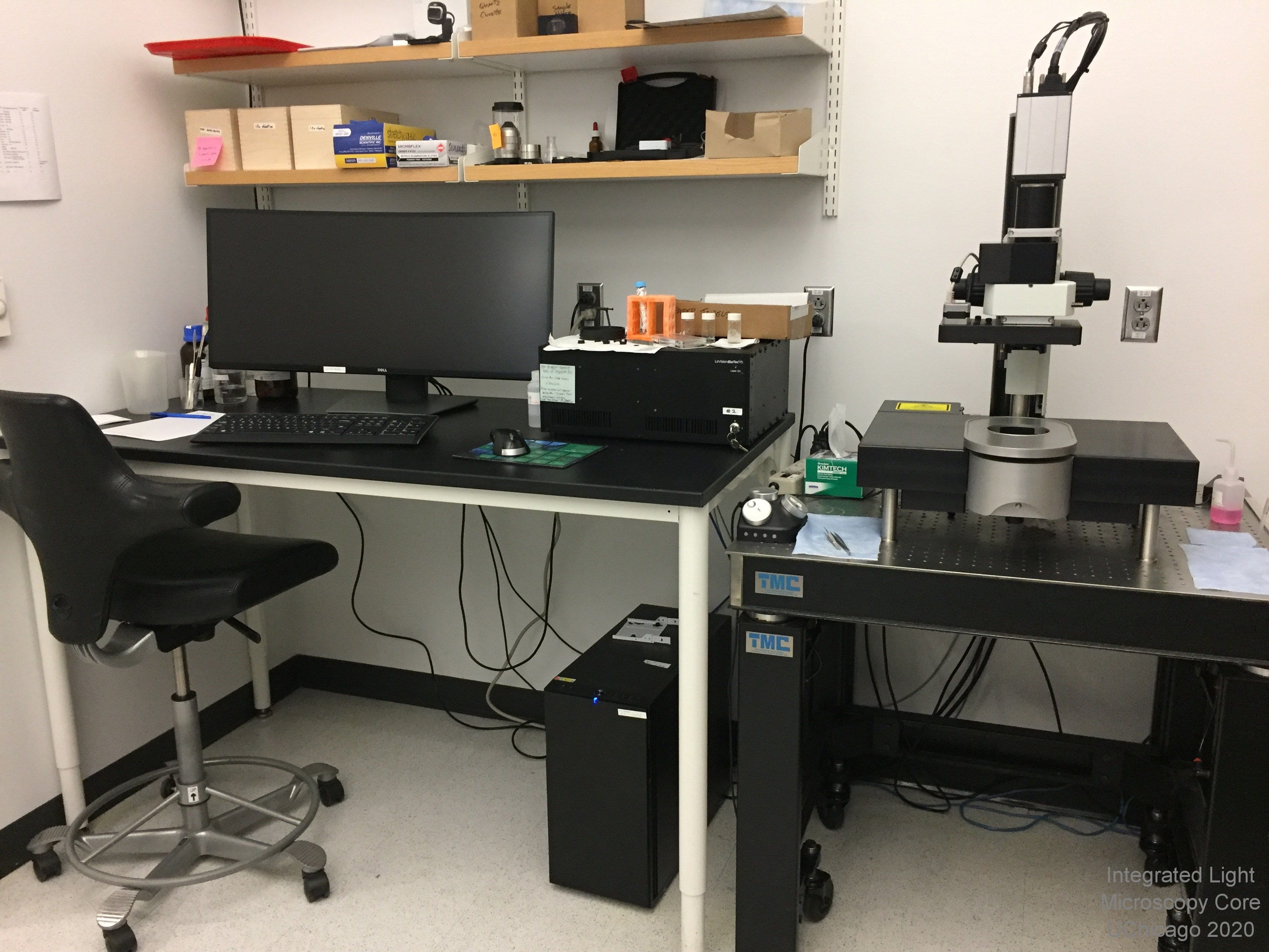 Overview: The LaVision Ultramicroscope is an upright lightsheet microscope ideal for large, fixed and cleared fluorescent samples. For comparisons of clearing techniques, Visikol has a nice chart detailing the basics of many commmon methods.
Location: KCBD 1250B
Training Contact: Christine Labno
Fluorophores this microscope can image:
- Cyan (CFP)
- Green (GFP, Alexa 488)
- Near Red (Alexa 543, Alexa 594)
- Far Red (Alexa 647)
- Near IR (Alexa 700)
Excitation Light Sources:
- solid state 440nm laser
- solid state 488nm laser
- solid state 561nm laser
- solid state 640nm laser
- solid state 785nm laser
Emission Detection and Filters:
- cyan emission filter at 480/40
- green emission filter at 525/50
- red emission filter at 620/60
- far red emission filter at 680/30
- near IR emission filter at 845/55
- Andor Zyla 4.2 megapixel sCMOS camera with 6.5um pixels
Objectives:
- 1.3x NA 0.08, 9mm working distance, dry
- 4x NA 0.3, 6mm WD, dipper with refractive index matching collar from 1.30 (water) to 1.55 (DBE)
- 12x NA 0.53, 10mm WD, dipper with two interchangeable RI-matching collars from 1.414-1.574 and 1.487-1.647
Sample Chamber:
- Upright format with dipping objectives
- Unique large, cuvette-based imaging chamber to allow for imaging of cleared specimens in clearing solution or other medium
- Inner dimensions of cuvette are 72 x 74 x 35mm
- Interchangeable sample holding platforms can accommodate samples up to 1cm^3
Special Features:
- Maximum lightsheet width = 17mm
- Total xy travel distance = 10mm
- Maximum scan area (xy) = 27mm^2 in a single field of view
- XYZ tiling imaging with Python-based stitching (Terastitcher) for mosaic-based imaging of large specimens
3i Lattice Lightsheet Microscope with Bessel Beam Illumination
 Overview: This is a commerically-produced clone of the Betzig system described in Science October 2014 and currently running at the HHMI Janelia Campus's Advanced Imaging Center. It is designed for low-light 3D time lapse imaging. This is NOT like the Zeiss Lightsheet.Z1 meant for large preps! Imaging is by means of water-immersion objectives and is limited to less than 100 micrometers depth of penetration. All samples MUST be on 5mm round coverslips. Please be sure to arrange all sessions in advance with Core Staff as use of this microscope requires careful calibration of the system before each use.
Location: KCBD 1250B
Training Contact: Christine Labno
Fluorophores this microscope can image:
- Cyan (CFP)
- Green (GFP, Alexa 488)
- Near Red (mCherry, Alexa 594)
- Far Red (Cy5, Alexa 647)
Excitation Light Sources:
- solid state 405nm laser
- solid state 488nm laser
- solid state 561nm laser
- solid state 642nm laser
- LED for brightfield illumination
Emission Detection:
- Hamamatsu Flash4v2+ sCMOS high-sensitivity, low noise camera (100nm pixel size)
Objective:
- Nikon 25x NA 1.1 water immersion objective
Sample Chamber:
- Due to sample holder configuration all samples MUST be on 5mm round #1.5 glass coverslips
- Heated bath chamber for live cell work. Perfusion is not practical due to large chamber volume
Features:
- High speed, low light 3D timelapse imaging.
- 100nm pixel size (xy)