
Live Sample Imaging
Environmental control for imaging at many temporal scales Check Microscope StatusNew User TrainingLive Sample Imaging
The microscopes in this listing are capable of modulating fluorescence excitation to preserve sample health. All have incubation chambers capable of temperature and humidity regulation; some also include CO2.
 There are several different categories of microscopy and many different spatial and temporal scales represented. We have systems capable of imaging multiple cell volumes per second, systems which image many fields of view over the course of several days, and systems which image whole organisms for several hours.
If you need help choosing the best microscope for your live sample work, contact Christine Labno with a description of your experiment and she can help you choose.
Hands-on Microscopes
To access a microscope, click the New User Training button above and work through our training checklist. Trained users have 24/7 access to the facility and are given permission to schedule their own microscope sessions though our online scheduling software.
SoRa Subdiffraction Marianas Spinning Disk Confocal - NEW February 2024!

Overview: Fully automated, inverted, Yokogawa-type spinning disk confocal ideal for imaging live samples. Features an automated XY stage and piezo-controled fine Z stage (focus) positioning for multi-point scanning plus incubator box for temperature, CO2 and humidity control. Slidebook software controls filter cube turret, objective turret, and high-speed brightfield shutter. SoRa with microvolution for nearly instantaneous deconvolution is ideal for subdiffraction (2-fold improvement over confocal) live cell imaging with little phototoxicity.
Location: KCBD 1250F
Training Contact: Khalil Rodriguez
Fluorophores this microscope can image:
- Violet/near UV (ex: DAPI, Alexa 405)
- Cyan (ex: CFP)
- Green (ex: GFP, Alexa 488)
- Yellow (ex: mCitrine, YFP)
- Orange (ex: Cy3, Alexa 543)
- Near Red (ex: Texas Red, Alexa 594)
- Far Red (ex: Cy5, Alexa 647)
Excitation Light Source(s):
- Near UV laser at 405nm
- Solid state laser at 445nm
- Solid state laser at 488nm
- Solid state laser at 561nm
- Solid state laser at 638nm
Emission Detection:
- Two Prime 95B Back Illuminated Scientific CMOS cameras (11x11um square pixels @ 18.7mm field of view)
- blue (DAPI) band pass emission filter at 445/58
- cyan (CFP) band pass emission filter at 482/35
- green (FITC, GFP) band pass emission filter at 525/50
- red (TRITC, mCherry) band pass emission filter at 600/50
- far red (Cy5) band pass emission filter at 706/95
Objectives:
- 10x / NA 0.3 dry (Olympus EC Plan Neofluar) working distance: 5.2mm
- 20x / NA 0.5 dry (Plan NeoFluar Ph2) working distance: 2.0mm
- 40x / NA 1.3 oil (Plan NeoFluar) working distance: 0.20mm
- 63x / NA 1.4 oil (Plan Apochromat) working distance: 0.14mm
- 100x / NA 1.3 oil (Plan NeoFluar) working distance: 0.20mm
Sample Chamber:
- Inverted platform for imaging of slides or dishes
- OKO full environmental control chamber (constant temperature, humidity and CO2)
- Motorized XY stage for multipoint timelapse and tiling
- Piezo controlled fine Z-stage positioning for 3D imaging
Special Features:
- Super-Resolution through Optical Reassignmnet (SoRa) is ideal for super-resolution live cell imaging and low phototoxicity. Specifications of SORa imaging plus deconvolution give approximately 2 fold increase in resolution (from 200nm to 100nm).
- CSU-W1 Yokogawa spinning disk allows for high speed imaging up to 200fps, wide field of view 16mm x 17mm, 25µm and 50µm pinhole disks for lower and higher magnification objectives
- Microvolution GPU accelerated Deconvolution Module for nearly instantaneous deconvolution.
- Options for split-view imaging, NIR imaging, illumination field flattening and super-resolution imaging
- Fast shutter speeds and channel switching for high speed imaging
- Vector high-speed point scanner for photoactivation/photoablation and FRAP experiments
- Full microscope automation through Slidebook software
Zeiss AxioObserver 7 automated, inverted widefield

Overview: Fully automated AxioObserver 7 widefield microscope with incubation chamber, fluorescence and DIC optics, full color histology, Prior motorized XY stage, and high NA objectives with auto-immersion capabilities at a wide range of magnifications and excellent Axiocam 705 monochrome and Axiocam 305 color CMOS cameras. Zeiss Zen software (blue edition) with AI automated sample finder, XYZT automation including time lapse, slide scanning, and mark and find multipoint over time capabilities. Fluorescence filters for DAPI, cyan, green, yellow, red, far red, and near IR plus DIC prisms for select objectives.
Location: KCBD 1250F
Training Contact: Lorraine Horwitz
Fluorophores this microscope can image:
- blue (e.g. DAPI)
- cyan (e.g. CFP)
- green (e.g. GFP/Alexa 488)
- yellow (e.g. YFP)
- red (e.g. DsRed, mCherry, Alexa 594)
- far red (e.g. Cy5, Alexa 647)
- near IR (e.g. cy7)
Excitation Filters:
- 385/30
- 436/20
- 469/38
- 500/25
- 555/30
- 631/33
- 735/40
Emission Filters:
- 425/30
- 480/40
- 514/30
- 535/30
- 592/25
- 681/45
- 788/38
Excitation Light Source:
- LED light source
Emission Detection:
- Monochrome and color CMOS cameras
Objectives:
- EC Plan-Neofluar 5x dry/ NA 0.16/ FWD=18.5mm
- EC Plan-Neofluar 10x dry/ NA 0.3/WD=5.2mm
- Plan-Apochromat 20x dry/ NA 0.8/ FWD=0.55mm
- C-Apochromat 40x / NA 1.1 water - rewatering feature with correction color/ FWD=0.62mm at CG=0.17mm
- Plan-Apochromat 63x / NA 1.4 oil/ FWD=0.19mm
Sample Chamber: XYZ automated inverted microscope stage
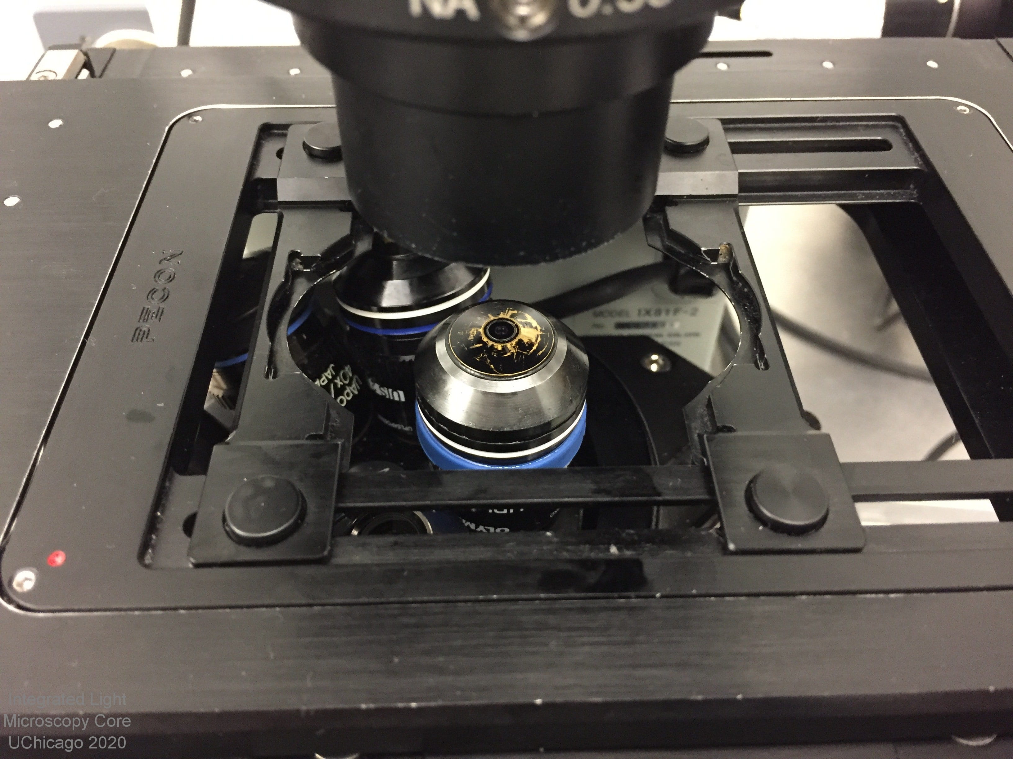
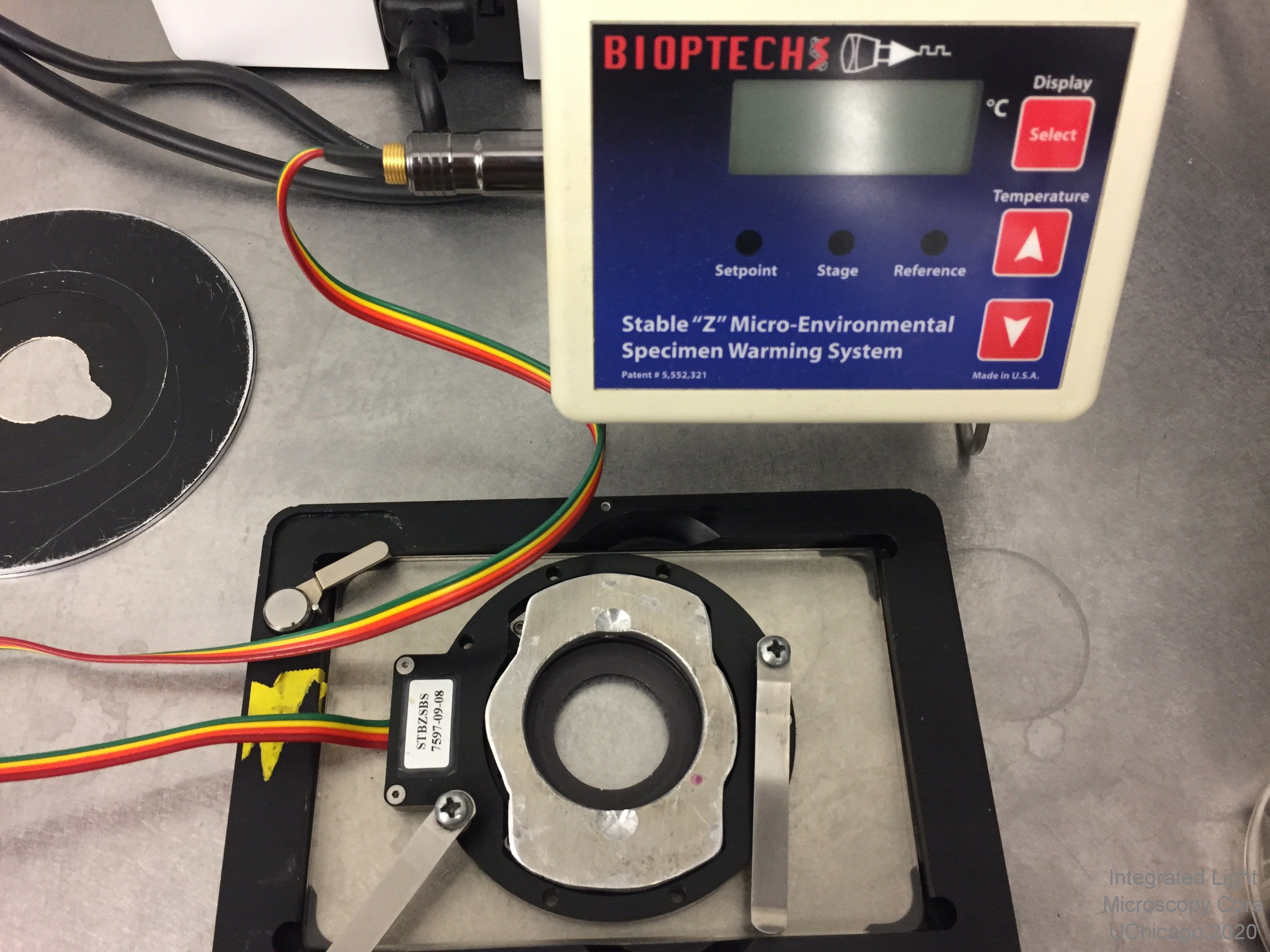
Olympus IX81 inverted widefield microscope
 Overview: This is an inverted, automated fluorescence microscope with a motorized stage and full-wrap incubation chamber. It is well suited to multi-well plate imaging, fixed cell fluorescence imaging, live cell imaging with real-time pixel intensity readouts, FRET experiments, ratiometric imaging (such as fura-2 340/380), and checking transfection efficiency.
Location: KCBD 1250C
Training Contact: Lorraine Horwitz
Fluorophores this microscope can image:
- Blue (e.g. DAPI)
- Cyan (e.g. CFP)
- Green (e.g. GFP, Alexa 488) -- 2 filter sets
- Yellow (e.g. YFP, mCitrine)
- Red (e.g. mCherry, Alexa 543) -- 2 filter sets
- Far red (e.g. Cy5, Alexa 647) -- 2 filter sets
- Near IR (e.g. Cy7)
- Fura-2 ratiometric Ca++ dye
Excitation and Emission filters:
- penta (5 color) dichroic with narrow filters / fast switching - blue ex: 387/11 em: 440/40 dichroic: 408; greens ex: 485/20 HQ em: 525/32 dichroic: 504; reds ex: 560/25 em: 650/13 dichroic: 581; far reds ex: 607/36 em: 684/24 dichroic 667; near IR ex: 740/13 em: 809/81 dichroic 762
- CFP ex: 436/20 and em: 480/40 (must be installed prior to use)
- green single cube - ex: 480/40 HQ and em: 510LP
- YFP ex: 500/20 and em: 535/30HQ (must be installed prior to use)
- red single cube - ex: 530-550 and em: 590LP
- far reds (Cy5) single cube - ex: 620/60 HQ and em: 700/75 HQ
- Fura-2 ex: 340; 380 and em: 510/40
- Chameleons 2 ex: 440/20 and em: 535/35; 485/40 (must be installed prior to use)
Excitation Light Source: Mercury arclamp
Emission Detection: Hamamatsu Orca Flash 4.0 camera with 4 megapixels (6.5um pixel size, 2048 x 2048 pixels). Huge field of view, great for tissue samples and multi-well plates
Objectives:
- 4x / 0.16 dry
- 10x / NA 0.3 dry (UPlanFL)
- 20x / 0.4 dry (LC PlanFL)
- 40x / 1.35 oil UV (UApo/340, best for FURA)
- 60x / 1.45 oil (PlanApo, TIRF rated)
- 100x 1.45 oil (TIRF rated)
- Also available (must be swapped in):
- 2x / 0.06 dry
- 40x / 0.6 dry LWD
- 150x / 1.45 oil (TIRF rated)
Sample Chamber:
- Automated, inverted platform (model IX81) for imaging on slides, live cell dishes or multi-well plates
- Full-wrap incubator box with micro CO2 chamber available
- Marzhauzer xy motorized stage for precise control of tiling, multi-point sampling or multi-well plate reading
- Chamber slide users see our chamber slide use warning before starting your cultures!
Special Features:
- SlideBook imaging software image capture in 2D, 3D, time lapse, multipoint, tiling, multi-well or any combination
- Zero Drift Correction (TM) auto re-focusing system. Especially useful for long term time lapse and / or heated preps
- DIC polarizer/analyzer plus appropriate prisms for most objectives
- Fura-2 340/380 ratiometric imaging
- CFP/YFP and Chameleon FRET filter set
- Automated filter changer with single color filters and a penta dichoic / filters for fast multi-color fluorescence imaging
- A beam splitter is available, but not currently installed. Be aware there is significant vignetting due to the large chip size of the Flash 4.0 camera. The beam splitter is for simultaneous capture of two emission wavelengths with the SAME excitation wavelength. Splitters available are: green/red (520/30 and 630/50) and blue/yellow-orange (460/50 and 570/60).
Olympus "live cell" DSU Spinning Disk Confocal
 Overview: The Olympus "live cell" DSU can be used for either widefield or confocal fluorescence capture, and since the entire field of view is illuminated at once, z-stack and time lapse capture can be faster than on the laser scanning confocals. The live cell DSU is on an inverted platform and features water immersion objectives, a motorized stage and filters for a broad range of fluorophores, from blue to far red, plus DIC optics.
Location: KCBD 1250F
Training Contact: Christine Labno
Fluorophores this microscope can image:
- Violet/near UV (ex: DAPI, Alexa 405)
- Cyan (ex: CFP)
- Green (ex: GFP, Alexa 488)
- Yellow (ex: mCitrine, YFP)
- Orange (ex: Cy3, Alexa 543)
- Near Red (ex: Texas Red, Alexa 594)
- Far Red (ex: Cy5, Alexa 647)
Excitation Light Source and Filters:
- Lumencore SOLA Fish LED light engine for even, flicker-free illumination
- blue/violet excitation filter at 350/50
- blue excitation filter at 405/12
- cyan excitation filter at 436/10
- green excitation filters at 480/25 and 490/20
- yellow excitation filter at 500/20
- orange excitation filter at 555/28
- red excitation filter at 565/25
- far red excitation filter at 635/20
Emission Detection:
- Photometrics Evolve- EMCCD camera (512 x 512, 16um pixels)
- blue emission filter at 457/50nm
- cyan emission filter at 470/30
- green emission filters at 525/40 and 528/38
- yellow emission filter at 535/30
- red emission filters at 617/73 and 620/60
- far red emission filter at 685/40
Objectives:
- 10x / NA 0.3 dry (UPlanFlN, 10mm WD)
- 10x / 0.4 water (UPlanApo)
- 20x / 0.7 water (UApo340, 0.35mm WD)
- 40x / 1.15 water (UApo340, 0.25mm WD)
- 60x / 1.2 water (UPlanSApo, 0.28mm WD)
- 100x / 1.45 oil TIRFM (PlanApo)
- 2x / 0.06 dry (must be installed prior to use)
- 4x / 0.16 dry (must be installed prior to use)
- 40x / 0.6 dry LWD (must be installed prior to use)
- 150x / 1.45 oil (must be installed prior to use)
 Sample Chamber:
- Fully automated, inverted platform (model IX81) for imaging on slides, chamber slides, 35mm dishes and multi-well plates (see our guidelines on use of chamber slides and well plates)
- Heated stage platform and micro chamber available for live cell preps (35mm dish configuration)
- Motorized xy-galvo stage for imaging multiple areas per dish and automated tiling of large specimens
- Chamber slide users see our chamber slide use warning before starting your cultures!
Special Features:
- Image capture through SlideBook software
- 3D and 4D (volume over time) rendering through SlideBook
- 100% - 1.5% neutral density filters available
- Interchangable pinhole disks for many magnifications (special request only)
- Dual cube viewing through the occulars (blue/green, green/red and red/far red)
Leica SP5 2-photon Laser Scanning Confocal - 2-PHOTON LASER UNAVAILABLE
 NOTES: The chiller for the MaiTai multiphoton laser has FAILED therefore the MaiTai 2-Photon laser is currently out of service. The rest of the Leica SP5 2-photon microscope is working normally for now, so if your project does not require excitation at longer wavelengths (700nm+) it can continue. This includes intravital imaging without the multiphoton laser.
Leica swapped the Argon laser with the one on the SP5 STED. We recommend keeping the power level low (between standby and 20% under configurations->laser) to prolong the life of the laser.
The HyD2 detector is failing, signal detected is very low. We recommend avoiding this detector if possible.
Overview: This laser scanning confocal system features software selectable conventional or high-speed resonance scanner galvanometer system with three (chilled, high sensitivity) internal PMT detectors (spectral) and two external (NDD) PMT detectors. Six visible laser lines and a tunable NIR pulsed laser (Spectra Physics Mai Tai broadband 710-990nm) provide excitation. The system features turnkey operation and full software control in addition to the features listed below.
Location: KCBD 1250B
Training Contact: Lorraine Horwitz
Fluorophores this microscope can image:
- Blue (ex: DAPI, Alexa 405)
- Cyan (ex: CFP)
- Green (ex: GFP, Alexa 488)
- Yellow (ex: mCitrine, YFP)
- Orange (ex: Cy3, Alexa 543)
- Near Red (ex: Texas Red, Alexa 594)
- Far Red (ex: Cy5, Alexa 647)
- Near IR (possible but not ideal, ex: Alexa 700)
- 2-photon imaging of Blue, Cyan, Green, Yellow, Orange - NO LONGER IN SERVICE
Excitation Light Source(s):
- near UV laser at 405nm
- 5 line Argon laser at 458, 476, 488, 496 and 514nm
- HeNe laser at 561nm
- HeNe laser at 594nm
- HeNe laser at 633nm
- MaiTai laser tunable from 710 - 990nm - NO LONGER IN SERVICE
Emission Detection:
- Custom emission detection of any wavelength range between 410nm and 800nm
- 3 chilled photon multiplier tubes (PMTs)
- 2 hybrid GaAsP/PMT detectors (HyDs)
- 2 non-descanned detectors (NDDs)
- Transmitted light detector
- 8-, 12-, or 16-bit grayscale output
Objectives (with additional optical zoom possible):
- 20x / NA 0.8 dry
- 40x / 1.25-0.75 oil
- 63x / 0.9 water
- 63x / 1.4 oil
- 100x / 1.46 oil
- 25x / NA 0.95 water
- Objectives listed under the SP5 STED description (must be installed)
Sample Chamber:
- Custom fit full incubation jacket (clear) for increased thermal stability during live cell experiments
- Inverted platform for imaging of slides or dishes
- LSM objective inverter device (allows system to be used as if it were an upright microscope platform)
- Automated XY stage for tiling / multipoint scanning
- "SuperZ" galvo focusing stage, 1.5 mm range. Demonstrated capability of three cell volumes per second (12 x 1-mm optical slices each)
- Chamber slide users see our chamber slide use warning before starting your cultures!
Special Features:
- FRET, deconvolution, and 3D software wizards
- Automated notch filters
- Spectra Physics Mai Tai broadband Ti:sapphire laser with 10W pump (tunable from 710 to 990 nm) (up to 2W output at 810 nm) - NO LONGER IN SERVICE
- Dual scanning galvanometer mirrors (max 16,000 Hz scan rate) 25 images/sec at 512x512; strip scans to 333 fps (4 channels). Standard scanner with beam park, bleaching, imaging to 8k x 8k (64 megapixels) per channel.
- AOTF control of visible laser lines, EOM and ND filter attenuation of NIR
- Excitation and emission scanning capability with linear unmixing
- Automatically optimized confocal pinhole apertures
- Transmitted detector with LWD and oil lenses and filters suited for SHG imaging
Leica SP5 STED Laser Scanning Confocal - STED AND ARGON LASER UNAVAILABLE
 The Leica SP5 STED was purchased with funds from NIH S10 OD010649 granted to Dr. Gopal Thinkaran and Dr. Vytas Bindokas in 2012
NOTE: The 592 depletion laser, and the Argon laser (458, 476, 488, 496, 514nm) on this system are no longer functional. We do not have a service contract for this end-of-life system, so we will not be replacing these lasers. Anyone in need of confocal imaging with DAPI can use the SP5 2-photon, SP8, or Stellaris. Anyone in need of super-resolution imaging has likely already moved to the SP8 3 color 3D STED system. The rest of the system continues to function, so confocal with red (561 and 594nm) and far red fluorophores (633nm) is still possible.
Leica SP5 STED - May 21, 2025 - We swapped this Argon (488nm) laser with the one from the SP5 2-photon which was starting to malfunction. The Argon laser is no longer functional and there is no service contract for this end-of-life system so we may have to retire this system by the end of the year.
Overview: The SP5 II is an advanced, high speed laser scanning confocal platform. It includes an acousto-optical beam splitter (AOBS) to select/introduce most excitation laser lines. There are eight excitation lines available, spanning the spectrum from green to far red. The acousto-optical tunable filters (AOTF) make it possible to detect a wide range of emission wavelengths with unlimited range on each of three PMTs (photo multiplier tubes), two HyDs (hybrid GaAsP detector) or two APDs (avalanche photodiodes).
Location: KCBD 1250F
Training Contact: Lorraine Horwitz
Fluorophores this microscope can image:
- Cyan (ex: CFP)
- Green (ex: GFP, Alexa 488)
- Yellow (ex: mCitrine, YFP)
- Orange (ex: Cy3, Alexa 543)
- Near Red (ex: Texas Red, Alexa 594)
- Far Red (ex: Cy5, Alexa 647)
- Near IR (possible but not ideal, ex: Alexa 700)
Excitation Light Source(s):
- 5 line Argon laser at 458, 476, 488, 496, and 514nm (unavailable)
- DPSS laser at 561nm
- orange HeNe laser at 594nm
- red HeNe laser at 633nm
Emission Detection:
- Custom emission detection of any wavelength range between 410nm and 800nm
- 3 chilled photon multiplier tubes (PMTs)
- 2 hybrid GaAsP/PMT detectors (HyDs) with optional time gating
- 2 internal avalanche photodiode detectors (APDs) for high sensitivity (green/red and CFP/YFP filters available)
- Transmitted light detector with DIC polarizer/analyzer plus prisms for most objectives available
- 8-, 12-, or 16-bit grayscale output
Objectives (base magnification, additional optical zoom is possible):
- 10x / NA 0.4 dry wd=2.74mm
- 20x / 0.7 multi-immersion (water, oil or glycerol) wd=0.26-0.17mm
- 40x / 1.25-0.75 oil wd=0.22mm
- 63x / 1.4-0.6 UV oil wd=0.14mm
- 100x / 1.40 oil (STED-rated) wd=0.13mm
- 50x / 0.9 dry wd=0.28mm (must be installed prior to use)
- 63x / 1.3 glycerol CORR wd=0.30mm (must be installed prior to use)
Sample Chamber:
- Full wrap incubator box with warm air heating for live samples
- Inverted platform for imaging of slides or dishes
- Automated XY stage for tiling / multipoint scanning
- "SuperZ" galvo focusing stage, 1.5 mm range. Demonstrated capability of three cell volumes per second (12 x 1-mm optical slices each)
- Chamber slide users see our chamber slide use warning before starting your cultures!
Special Features:
- Continuous wave depletion laser at 592nm allows for single or dual color super resolution (STED method) with either the resonant (high speed) or galvo (high pixel density) scanners.
- STED mode allows for resolution of particles down to 50nm FWHM (cyan, green, yellow fluorophores only)
- Tandem scanner with dual scanning galvanometer mirrors allows for either high speed scanning (max 16,000 Hz scan rate) 25 images/sec at 512x512; strip scans to 333 fps (5 channels) OR high pixel density scanning (imaging to 8k x 8k [64 megapixels] per channel).
- Standard scanner includes beam park for FRAP, bleaching and photoactivaiton
- Sequential scanning capability allows for rapid sequential scanning of fluorophores with minimal bleed-through or cross-talk
- Wizards for FRAP and FRET
- AOTF (acousto-optical tunable filters) for spectral scanning, allowing separation of fluorophores with similar ranges (e.g. GFP and FITC) through spectral unmixing
- LAS_AF Leica confocal software on Windows
- Off-line version of LAS_AF software available on facility workstation
3i Lattice Lightsheet Microscope with Bessel Beam Illumination
 Overview: This is a commerically-produced clone of the Betzig system described in Science October 2014 and currently running at the HHMI Janelia Campus's Advanced Imaging Center. It is designed for low-light 3D time lapse imaging. This is NOT like the Zeiss Lightsheet.Z1 meant for large preps! Imaging is by means of water-immersion objectives and is limited to less than 100 micrometers depth of penetration. All samples MUST be on 5mm round coverslips. Please be sure to arrange all sessions in advance with Core Staff as use of this microscope requires careful calibration of the system before each use.
Location: KCBD 1250B
Training Contact: Christine Labno
Fluorophores this microscope can image:
- Cyan (CFP)
- Green (GFP, Alexa 488)
- Near Red (mCherry, Alexa 594)
- Far Red (Cy5, Alexa 647)
Excitation Light Sources:
- solid state 405nm laser
- solid state 488nm laser
- solid state 561nm laser
- solid state 642nm laser
- LED for brightfield illumination
Emission Detection:
- Hamamatsu Flash4v2+ sCMOS high-sensitivity, low noise camera (100nm pixel size)
Objective:
- Nikon 25x NA 1.1 water immersion objective
Sample Chamber:
- Due to sample holder configuration all samples MUST be on 5mm round #1.5 glass coverslips
- Heated bath chamber for live cell work. Perfusion is not practical due to large chamber volume
Features:
- High speed, low light 3D timelapse imaging.
- 100nm pixel size (xy)
Olympus VivaView in-incubator microscope - DECOMMISSIONED FEB 2024
 This microscope is no longer available for use, it has been replaced with a Zeiss Axio Observer 7. The information here is for publication generation purposes only.
Overview: The Olympus LCV110U VivaView is an innovative system for long-term, multi-dimentional, live cell imaging. The system is an inverted widefield fluorescence microscope built into an incubator box. The incubator keeps optimal temperature, humidity and CO2 during long experiments and decreases the effects of thermal drift and evaporation. Allows for imaging of multiple areas in up to eight dishes, kept at 37C, 5% CO2. Brightfield, blue (DAPI), cyan (CFP), green (GFP) and near red (mCherry, DsRed) imaging available.
Location: This microscope is no longer available for use
Training Contact: N/A
Fluorophores this microscope can image:
- blue (e.g. DAPI)
- cyan (e.g. CFP)
- greens (e.g GFP, Alexa 488)
- reds (e.g. mCherry, Alexa 543)
- far reds (e.g. Cy5, Alexa 633)
Excitation Light Source and Filters:
- X-Cite eXactate arclamp light source with liquid light guide can be adjusted to lower light intensity and minimize photobleaching.
- blue excitation filter at 387/11nm; dichroic 409nm
- cyan excitation filter at 436/20; dichroic 445
- green excitation filter at 470-495; dichroic 505
- red excitation filter at 530-550; dichroic 570
- far red excitation filter at 620/60; dichroic 700
Emission Detection and Filters:
- Air-cooled (-35C) CCD camera gives 12-bit grayscale images with 1344 x 1024 pixels (6.45 micron pixels)
- blue emission filter at 447/60
- cyan emission filter at 480/40
- green emission filter at 510-550
- red emission filter at 575IF (long pass)
- far red emission filter at 700/75
Objective:
- 40x / NA 0.95 dry
- 0.5x and 2x magnification changers create 20x and 80x magnification
Sample Chamber:
- Fully enclosed, humidified incubator set to 37C, 5% CO2.
- Inverted platform with 8 position motorized carousel for imaging multiple areas across multiple 35mm round live-cell dishes (#1.5 glass bottom dishes recommended, MatTek dishes are available in sleeves of 10 in the facility)
Special Features:
- Independent objective and carousel heaters minimize thermal drift
- MetaMorph image capture software
- Data are saved to hard drive immediately post-capture
- Optional motorized z-stack collection for 3D or focus correction
Zeiss Lightsheet Z.1 - Moved to OBA CDIF April 2022
The Zeiss Lightsheet Z.1 was provided by the Institute for Genomics & Systems Biology (IGSB).
 This microscope is no longer available through the Integrated Microscopy Core. As of April 2022 it is housed and managed by the Organismal Biology and Anatomy CDIF Facility.
Overview: The Zeiss Lightsheet Z.1 is one of the first commercially available Single Plane Illumination microscopes. It is particularly suited for temperature controlled, long-term imaging of living samples such as zebrafish or Drosophila embryos. Samples can be kept in the chamber and imaged for hours or days. Sensitive cameras and single plane illumination allow for gentle sample illumination, reducing photodamage.
Location: Moved to Oganismal Biology and Anatomy CDIF Facility April 2022
Training Contact: Oganismal Biology and Anatomy CDIF Facility
Fluorophores this microscope can image:
- Cyan (CFP)
- Green (GFP, Alexa 488)
- Near Red (DsRed, mCherry)
Excitation Light Sources:
- solid state 445nm laser
- solid state 488nm laser
- solid state 561nm laser
Emission Detection and Filters:
- Two chilled PCO-Edge scMOS 16-bit cameras with 1920 x 1920 pixels for fast, detailed, simultaneous two color imaging.
- BP 460-500nm (CFP),
- BP 505-545nm (GFP/YFP),
- SP 550nm (GFP/YFP),
- LP 585nm (mCherry)
Objectives:
- 5x / NA 0.16 dry (uses 5x NA 0.1 dry illumination objectives)
- 20x / 1.0 water dipper (uses 10x NA 0.2 dry illumination objectives)
- 40x / 1.0 water dipper (uses 10x NA 0.2 dry illumination objectives)
Sample Chamber:
- Sample is held in a buffer-filled chamber during imaging. PBS, seawater and nutrient buffers (e.g. E3) have all been used successfully.
- Water cooled/heated Peltier block for chamber temperature settings from 10C-42C. Temperature feeds back to the software from both the block and the sample chamber.
Special Features:
- Green/red or cyan/red simultaneous imaging is possible. Cyan/green or three color serial imaging is also possible.
- 360 degree sample rotation plus programming for imaging from multiple angles. Incorporation of beads allows for multi-angle 3D reconstructions.
- Off-line version of Zeiss Zen software available on facility workstation.