
Drop Off Service
We have two slide scanners that are run purely as a drop-off service.
To use the service: first see our Slide Preparation and Practical Considerations documents.
First-time Core users, please submit a Facility User Record and make sure you can access the OSRF FTP server for data retrieval (Don't have access? Use this form to request!).
All users need to bring a completed copy of the Scanner Service Request Form and your slides in a container labeled with your name to KCBD 1250
NOTE ON CLINICAL SAMPLES - All slides containing patient samples must be de-identified before scanning (i.e. protected health information must be covered, blacked out or otherwise removed)
We will add your slides to the queue and email you when your scans are done. Scan files will be available for download on the OSRF FTP server. Physical slides should be picked up from designated areas as soon as possible after scanning. Slides not picked up within 3 months become the property of the Core and may be discarded if we run out of storage space.
To view your images: For CRi .mrxs files use CaseViewer (PC only) or QuPath; for Olympus VS200 .vsi files use OlyVIA or QuPath. We have created guides for basic OlyVIA and basic QuPath use
Contact Khalil Rodriguez for more information on this service
Olympus VS200 Slideview Research Slide Scanner
Click for freeOlyVIA software download or free QuPath whole slide image analysis software from the University of Edinburgh
The Core has created a guide for beginning OlyVIA software use (in progress) and a guide for beginning QuPath use with whole slide scans. The QuPath developers have extensive documentation as well.
Click for our Slide Preparation Guidelines and our Practical Considerations for Slide Scanning
Overview: Slide scanning on the Olympus VS200 Slideview is run as a drop off service. Our whole slide scanners create a virtual library of histology or multi-channel fluorescent slides. Details on the virtual slide can then be zoomed to a virtual magnifications from 0.5x-100x and exported as .tif or .jpeg for analysis or presentation with other software packages. We currently recommend processing with either the opensource QuPath software package or the proprietary free download from Olympus called OlyVIA.
Location: KCBD 1250E
Drop off Contact: Khalil Rodriguez
This microscope can image:
- Full color histology imaging with white light
- Blue (DAPI) ex 378/52 em 432/36 (Quad dichroic)
- Cyan (CFP) ex 438/24 em 482/25 (Triple dichroic)
- Green (FITC, GFP) ex 474/27 em 515/30 (Quad dichroic)
- Yellow (YFP) ex 509/22 em 544/24 (Triple dichroic)
- Orange (TRITC) ex 554/23 em 595/31 (Quad dichroic)
- Red (mCherry) ex 578/21 em 641/75 (Triple dichroic)
- Far red (Cy5) ex 635/18 em 698/70 (Quad dichroic)
- Near IR (Cy7) ex 735/28 em 809/100 (Single dichroic)
Excitation Light Sources:
- X-Cite Xylis broad spectrum LED from Encelitas
Emission Detection:
- Hamamatsu Orca Fusion digital camera for fluorescence (3.45um pixel size binned 2x2 for final 6.9um pixel size, 5.3 megapixels with 80% QE)
- Integrated 2/3 inch CMOS for histology (3.45 μm × 3.45 μm pixel size, high sensitivity, high resolution)
Objectives:
- 2x, 4x, 10x NA 0.4, 20x NA 0.8, and 40x NA0.95 dry
- 60x NA 1.42 oil (small regions only, file size and scan time can balloon quickly)
Pixel Sizes:
- 2x - 3.275 um/pixel (fluorescence), 2.7382 um/pixel (histology)
- 4x- 1.507 um/pixel (fluorescence), 1.3691 um/pixel (histology)
- 10x - 0.6504 um/pixel (fluorescence), 0.548 um/pixel (histology)
- 20x- 0.3251 um/pixel (fluorescence), 0.274 um/pixel (histology)
- 40x - 0.1626 um/pixel (fluorescence), 0.137 um/pixel (histology)
- 60x - 0.1084 um/pixel (fluorescence), 0.091 um/pixel (histology)
Special Features:
- Drop off service: drop off your slides and put your name in the queue. We'll do the rest and notify you when your scans are done. Slides are scanned on a first-come, first-served basis.
- Automated scanning of up to 210 slides per run, unlimited slides per batch
- Data is available on our server, so you can download it straight to any campus computer.
- Offline image viewing software available free from Olympus (Windows only) or analysis software free from QuPath at the University of Edinburgh (multi-platform: Windows, MacOS, Linux).
CRi Pannoramic MIDI 20x Whole Slide Scanner
The CRi scanners are now over a decade old and are no longer supported in terms of repairs, parts or even purchasing a new version of the system (no US distributor). They are starting to malfunction more frequently and while we are currently able to repair them in house, there may come a time when that is not possible. We will keep them running as long as we are able, but they may die without warning at any time. We are encouraging existing scanner clients to move their scans to the newer Olympus VS 200 as soon as they are comfortable. We're happy to scan the same slide in both machines so that you may compare scan quality.Click for free CaseViewer software download or free QuPath whole slide image analysis software from the University of Edinburgh
Click for our Slide Preparation Guidelines and our Practical Considerations for Slide Scanning
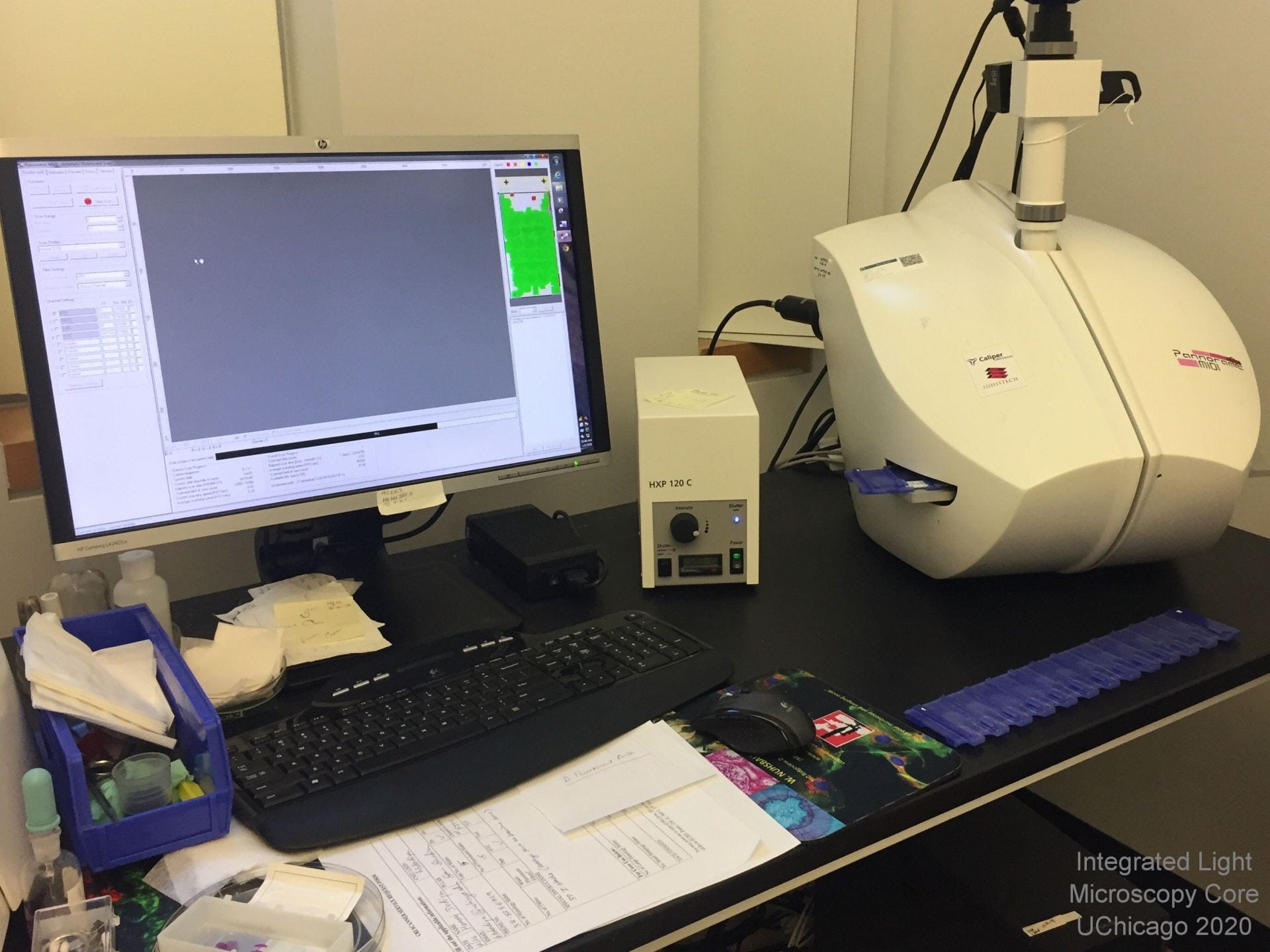 Overview: Slide scanning on the Pannoramic scanners is run as a drop off service. Our whole slide scanners from Cambrige Research and Instrumentation (CRi, now owned but NOT SUPPORTED by a spinoff of ThermoFisher) create a virtual library of histology or multi-channel fluorescent slides. Using the proprietary software available in the facility or as a free download, details on the virtual slide can then be zoomed to a virtual magnifications from 0.5x-100x and exported as .tif or .jpeg for analysis or presentation with other software packages.
Location: KCBD 1250E
Drop off Contact: Khalil Rodriguez
Fluorophores this microscope can image:
- Blue (e.g. DAPI)
- Green (e.g. GFP, Alexa 488)
- Red (e.g. mCherry, Alexa 594)
- Far Red (e.g. Alexa 647)
- Full color histology imaging with white light
Excitation Light Sources:
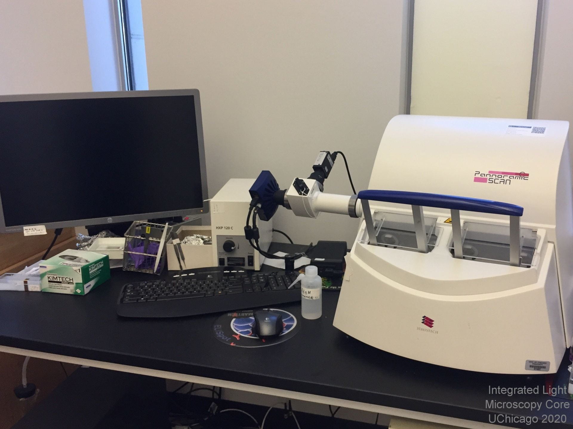
- HXP 120 C arclamp with fiber optic lightguide for fluorescence
- Incandescent white light bulb for histology
Emission Detection:
- Zeiss AxioCam MRm high sensitivity, 12-bit greyscale, 6.45um per pixel camera for fluorescence imaging
- CIS 3CCD 3 megapixel color camera for histology
Objective: 20x Zeiss LWD objective, virtual magnification range from 0.5x to 100x
Pixel Sizes: 0.3225 um/pixel (fluorescence) and 0.3225 um/pixel (histology)
Special Features:
- Drop off service: drop off your slides and put your name in the queue. We'll do the rest and notify you when your scans are done. Slides are scannned on a first-come, first-served basis.
- Automated scanning of up to 12 slides per batch
- Data is available on our server, so you can download it straight to any campus computer.
- Offline image viewing software available free from 3DHistech (Windows only) or analysis software free from QuPath at the University of Edinburgh (multi-platform: Windows, MacOS, Linux).
CRi Pannoramic SCAN 40x Whole Slide Scanner - NO LONGER FUNCTIONAL
The 40x CRi scanner has broken down after 13 years of service. There is no service contact and currently no US distributor, so we cannot repair or replace this system. This information is here for legacy purposes ONLY (grant and publication writing with existing scans). Please choose one of our other two scanners for your project.
Click for free CaseViewer software download or free QuPath whole slide image analysis software from the University of Edinburgh
Click for our Scanner Slide Preparation Guidelines and our Practical Considerations for Slide Scanning
Drop off Contact: No longer in service, please choose a different system
This microscope can image: nothing currently, it is broken and cannot be repaired. Information here is for legacy purposes only.
Emission Detection: Allied Vision Technologies Stingray F146C color, 4.6 um per pixel camera
Objective: 40x NA 0.95 LWD Zeiss objective, virtual magnification range from 0.5x to 100x on high resolution (down to 0.12um/pixel) tiled images
Pixel Sizes: 0.1644 um/pixel (fluorescence) and 0.1645 um/pixel (histology)
Special Features:
- Drop off service: drop off your slides and put your name in the queue. We'll do the rest and notify you when your scans are done. Slides are scanned on a first-come, first-served basis.
- Automated scanning of up to 150 slides per batch
- Data is available on our server, so you can download it straight to any campus computer.
- Offline image viewing software available free from 3DHistech (Windows only) or analysis software free from QuPath at the University of Edinburgh (multi-platform: Windows, MacOS, Linux).
Hands-on Microscopes
To access a microscope, click the New User Training button above and work through our training checklist. Trained users have 24/7 access to the facility and are given permission to schedule their own microscope sessions through our online scheduling software.
Caliber I.D. RS-G4 Large Format Laser Scanning Confocal
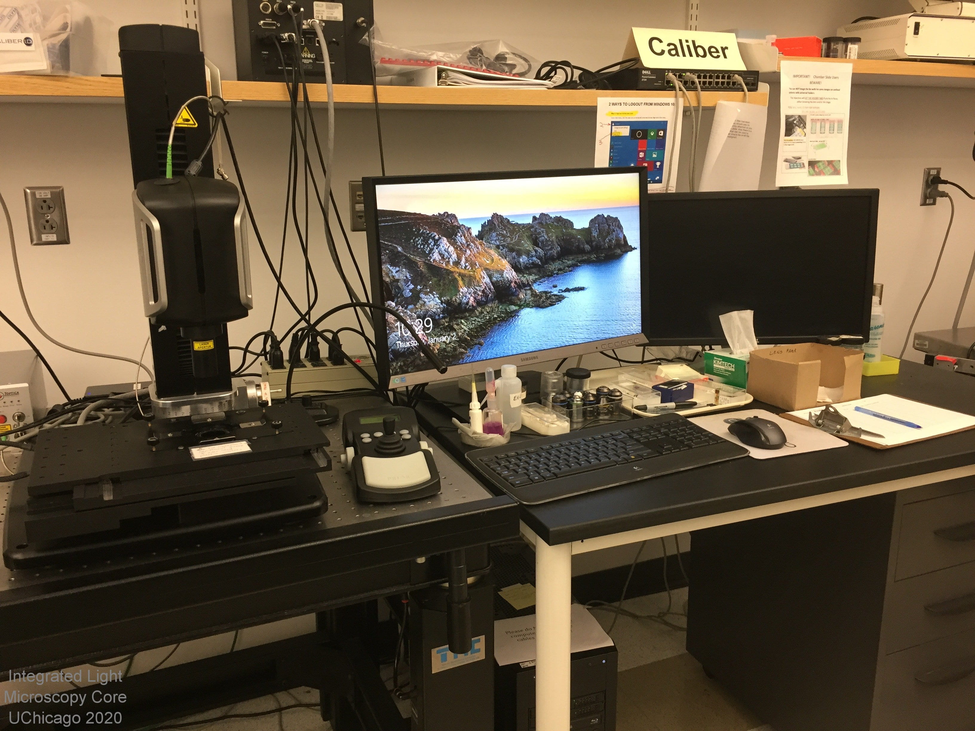 Overview: The Caliber I.D. RS-G4 is an upright laser scanning confocal microscope with a motorized XY-Z stage and software designed for large format (tiled) images. This system is ideal for high speed, high quality tiling in 2D or 3D and will handle samples up to 120mm x 80mm x 6mm thick.
Location: KCBD 1250F
Training Contact: Christine Labno
Fluorophores this microscope can image:
- Blue (DAPI, Alexa 405)
- Cyan (CFP)
- Green (GFP, Alexa 488)
- Yellow (mCitrine, YFP)
- Orange (Cy3, Alexa 543)
- Near Red (Texas Red, Alexa 594)
- Far Red (Cy5, Alexa 647)
- Near IR (Alexa 700)
Excitation light source(s):
- Near UV laser at 405nm
- solid state laser at 488nm
- solid state laser at 561nm
- solid state laser at 640nm
- solid state laser at 785nm
Emission detection:
- 2 photon multiplier tubes (PMTs) for emission detection
- Seven band pass filters for emission range selection: 450/70, 520/44, 550/88, 600/52, 630/69, 670/30 and 832/37
- 16-bit tif or Imaris .ims file output.
Objectives (base magnification, additional optical zoom is possible):
- Supports dry, oil and water immersion objectives with a wide range of magnifications available (2x - 100x).
- Objectives must be programmed in prior to use.
Sample Chamber:
- Upright platform for imaging of slides
- Imaging in dishes limited, only possible with dipping (coverslip-free) objectives
- Fast ribbon / strip mosaic scan mode plus conventional tiling mode, both with real time / online stitching
Special Features:
- 8kHz Resonant-scanning confocal
- Large mosaic creation up to 120 x 80 mm with high precision scanning stage.
- Fluorescence and/or reflectance confocal imaging
- Automated scanning of single, simultaneous pairs or sequential channels.
- Automated collection of time-lapse, X-Y-Z, multi-stage points, multiple wavelengths and snapshots.
- 1024 x 1024 pixel resolution