The Fehon Lab
Cellular organization and function of the Hippo Pathway in tissue growth controlImage Gallery

Yellow fluorescent protein (YFP) tagged Merlin, endogenously expressed, is highly localized at the apical domain of living wing imaginal disc cells, predominately at the junctional and medial cortex.
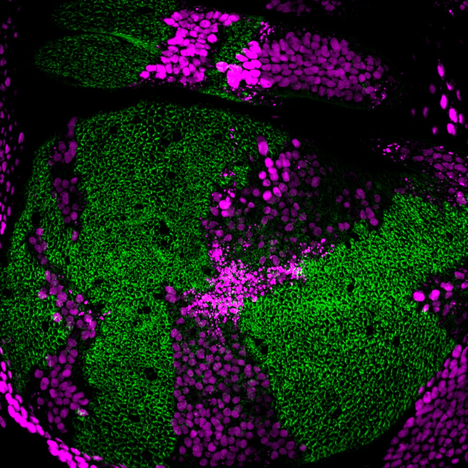
The abundance of Kibra protein (green) increases dramatically in somatic mosaic clones of Merlin mutant cells (marked by the absence of RFP-magenta) due to decreased Kibra degradation.

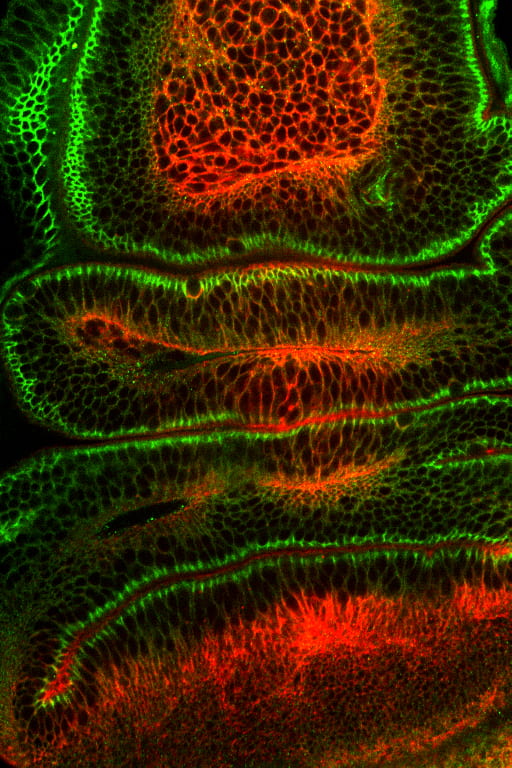
Optical section of a fold in the Drosophila wing epithelium stained for Moesin (red, apical membrane), E-cadherin (blue, adherens junctions) and Coracle (green, septate junctions).

A clone of wing imaginal disc cells (green) expressing dominant-negative Moesin crawling over the surface of the epithelium (epithelial marker red).

Drosophila larval salivary gland, a monolayered epithelial tube, expressing Merlin-GFP (green) and stained with propidium iodide (nuclei; red).
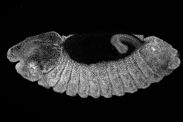
Drosophila embryo (dorsal closure stage) stained with an anti-Coracle antibody.

Pupal eye from Drosophila stained with antibodies for the Notch protein.
Optical section of the Drosophila wing epithelium stained for actin (red) and Coracle (green) a component of the septate junction.
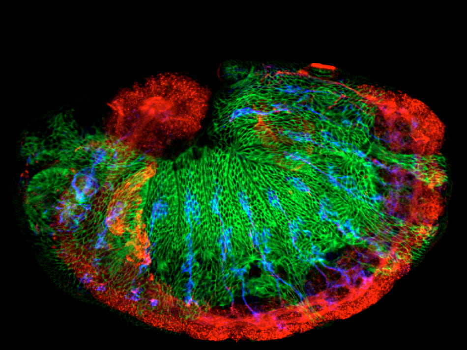
Drosophila Cdc42 mutant embryo, stained for Coracle (green), the peripheral nervous system (22C10, blue) and the central nervous system (anti-HRP, red).