With the recent addition of the NovoCyte Quanteon, the CAT Facility now employs instruments with three different classes of photodetector: Avalanche Photodiodes (APDs) on the Cytek Auroras, Silicon Photomultipliers (SiPMs) on the NovoCyte Quanteon and Penteon from Agilent, and the classic Photomultiplier Tubes (PMTs) on the BD LSR platforms and most everything else. In this blogpost I want to break down the role of a photodetector in flow cytometry, briefly describe how the different types work, and compare them based on their features relevant to flow.
Background
For a flow cytometer to be at all useful, it must convert the fluorescence of a labelled cell into discrete values that we can visualize on the classic bivariate plot. To drill down another level, this means a flow cytometer must be able to detect an input of photons, amplify it, and output an analog signal in the form of electric current (modern flow cytometers subsequently digitize that analog signal, but that’s beyond the scope of today’s blogpost). For example, say we want to compare the GFP expression of two reporter cell lines. The cells with more GFP will emit more green photons (fluorescence) which, once amplified, will lead to a proportionally greater electric current (signal).
This conversion is precisely what photodetectors are made for. There are many classes of photodetectors, and they accomplish this conversion is different ways, but today we’re focusing on the three in use by the CAT Facility’s instruments (APDs, SiPMs, and PMTs).
Why Do We Care?
Instrument manufacturers have their reasons for choosing a specific photodetector, but not all of them are relevant to the end user (that’s you!). A summary of how these photodetectors actually work is forthcoming, but we both know this section is why you clicked on the blogpost in the first place.
Usable nm Range
The photodetector types are most efficient at different wavelength ranges, with PMTs performing best in the 300-800nm range and SiPM/APDs winning out past the 700nm mark on up to 1000nm+.1 “Efficient,” in this case, means fewer photons failing to be detected. If your experiment relies on detecting the far-red signal of dim or hard-to-resolve populations, an SiPM/APD is your best bet. Running a multicolor panel with emission mostly on the lower end of the spectrum? Stick with PMTs.
As an aside, beware figures from manufacturers comparing photodetector efficiency over the spectrum, especially when the photodetector they use performs vastly better at all wavelengths. I find they’ve often cherry-picked the comparison to show an excellent example of their photodetector vs. a subpar example of “the other guy’s” photodetector.
Noise and Dark Counts
SiPM/APDs and PMTs have comparable (low) levels of noise, but again PMTs have an edge up until around 700nm. This is mostly due to “dark counts,” weak noise from thermal electrons that will increase with both an SiPM/APD’s temperature and the applied voltage. PMTs also experience dark counts, but at a much lower rate and, critically, they only increase with voltage. Thankfully, noise from dark counts (even from SiPM/APDs) typically show up at intensities on par with single-photon detection, so rarely is a dark count going to be misinterpreted as fluorescent signal.2 The larger impact is in the Scatter parameters (FSC and SSC) used to analyze morphology. Even low levels of noise can make it difficult to resolve small particles from debris. In such a case, you should take every advantage you can get and utilize a PMT-equipped instrument.
Dynamic Range
Dynamic range is the range of fluorescent values a cytometer can detect. More concretely, it’s the number of decades a cytometer can utilize on a logarithmic scale of fluorescence. Higher dynamic range means keeping brighter events on scale without losing resolution on the lower end. It will also make voltage optimization much easier, which is a selling point of the Quanteon and Penteon. On these NovoCyte platforms, the dynamic range is so large that it’s possible you won’t need to optimize the default voltages at all, as there will be a large range of possible voltages that keep your brightest events on scale while maintaining low-end resolution.
While it’s a critical feature of a flow cytometer, the value has more to do with the specific construction of the photodetector rather than PMTs broadly beating out SiPMs or vice versa. It’s not an exhaustive comparison, but see the table at the bottom of the page for a quick comparison of photodetector classes, including sample dynamic range values for relevant CAT Facility instruments.
Principles of Operation
As previously stated, all these photodetectors take photon input (fluorescence), amplify it, and output current (signal). However, the exact mechanism varies. If you’re not interested in the nitty-gritty, click here to skip to the next section for the comparison table and final thoughts.
 PMTs convert incoming photons to electrons, then accelerate and multiply those electrons successively to output a proportionally amplified current. They have three components: the photocathode, the dynode array, and the anode. The photocathode absorbs the incoming photons and emits primary electrons via the photoelectric effect. When these reach the first dynode, each primary electron gives rise to a cloud of new electrons via secondary emission. These new secondary electrons are all accelerated to the next dynode, where the process is repeated with ever-increasing dynode voltages, and an ever-increasing number of electrons. This chain reaction leads to a greatly multiplied number of electrons that, when striking the anode, will produce current proportional to the original photon input.
 APDs take incoming photons and spawn corresponding +/- electron pairs between oppositely charged poles, generating the output current from the resultant charge gradient. In their simplest form, APDs are a region of semiconductor (usually silicon) flanked by oppositely charged poles. When a photon strikes the semiconductor (called the depletion region) it generates a +/- electron pair via the photoelectric effect. These electrons move towards the pole of opposite charge, creating a charge gradient (current) to be measured as signal. In this case, the APD is in Linear Mode.

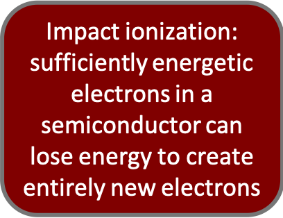
However, increasing the applied voltage to the APD will cause the electron pairs to accelerate that much faster towards the opposite pole. At a certain point (known as the breakdown voltage) the electron pairs move through the silicon so quickly that they generate entirely new electron pairs via impact ionization. Similarly to the function of the dynodes in the PMT, this leads to a greater amplification of the original photon input and greatly increases the gain. An APD operating in this manner is said to be in Geiger Mode.
SiPMs are simply arrays of APDs operating in Geiger Mode. Each APD is a part of a micro-cell, which includes a quenching resistor that quickly neutralizes the residual charge so the APD is quickly prepared to receive more photons. Having these in array and readily quenched means more sensitivity and a shorter refractory period in between detection events.
[Return to Principles]↩
| PMT | APD | SiPM | |
| Range (nm) | 300-800 | 400-1000+ | 400-1000+ |
| Internal Gain | 10^5-7 | 10^2 | 10^5-7 |
| Power Draw | Up to 1000V | 100-200V | Less than 100V |
| Dynamic Range | 5 Decades (Fortessa) | 6.5 Decades (Aurora)4 | 7.2 Decades (Quanteon)3 |
| Low Light Detection | Large active area and very rare dark counts | Smaller active area, more common dark counts (vs PMT) increasing with temperature | Array of microcells increases active area, more common dark counts (vs PMT) increasing with temperature |
| Noise | Relatively low until the ~800nm mark, increases with voltage and higher emission wavelengths | Noisier than PMT except at ~800nm+, but comparable over whole range | Noisier than PMT except at ~800nm+, but comparable over whole range |
Conclusion
There is no best photodetector for all experiments, but there is a best photodetector for every experiment! Consider what specific data you’re after, and from there you can decide which photodetector will best fit your needs. For a simple viability check, pretty much any flow cytometer in the core is sufficient. But if, for example, you’re running a 10 color panel wherein you need precise statistics on a very rare and very poorly resolved population emitting in the 600nm range, you’d probably want a PMT-equipped cytometer for its superior low light detection. On the other hand, if you’re expecting a huge range of possible brightnesses on a fluorophore, especially a red/far-red one, an SiPM-equipped cytometer would likely serve you best. Ease of use is another consideration. In the case of our new NovoCyte platforms, the super-high dynamic range means voltage optimization is potentially unnecessary. Considering suboptimal voltages are maybe the most common cause of suboptimal flow cytometry data, this can be a huge boon for user-friendliness.
Ultimately, you don’t have to decide by yourself! While this blogpost may be a useful starting point, CAT Facility Staff can always help guide you to the best instrument for your research.
1. [Lawrence WG, Varadi G, Entine G, Podniesinski E, Wallace PK. Enhanced red and near infrared detection in flow cytometry using avalanche photodiodes. Cytometry A. 2008 Aug;73(8):767-76. doi: 10.1002/cyto.a.20595. PMID: 18612992; PMCID: PMC2674288.]↩
2. [Patrick Eckert, Hans-Christian Schultz-Coulon, Wei Shen, Rainer Stamen, Alexander Tadday,
Characterisation studies of silicon photomultipliers, Nuclear Instruments and Methods in Physics Research Section A: Accelerators, Spectrometers, Detectors and Associated Equipment, Volume 620, Issues 2–3, 2010, Pages 217-226, ISSN 0168-9002, https://doi.org/10.1016/j.nima.2010.03.169.]↩
3. [Cytek Aurora Brochure]↩
4. [NovoCyte Quanteon Product Page]↩
Looking for more flow cytometry resources?
Sample Preparation
Learn about flow cytometry staining protocols, antibody titration, fixation considerations, etc.
Experiment Design
Learn about flow cytometry controls – FMOs vs isotype controls, cells vs compensation beads, etc.
Panel Design
Learn about the strategy and theory of flow cytometry panel design.


This post was very helpful in understanding the differences between traditional PMTs, APDs and SiPMs
Dear Bert, thanks for sharing your knowledge with us. This is a very good written article with valubale learnings about detectors in flow cytometry.
Let me please add something, regarding the the >800 nm wave length of PMTs, one known manufacturer uses special PMTs for better sensitivity > of 720nm signals, they use GaAs PMTs. Nevertheless I couldn’t find the wavelength reach of the used GaAs PMTs.
Thanks again