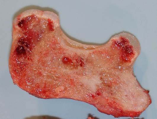
Osteochondromas are primary cartilaginous neoplasms that induce growth of underlying bone and marrow. These are usually benign, but cartilaginous overgrowth / thickness of cartilaginous cap may be a sign of malignant transformation.
Triage & Gross
- These specimens usually demonstrate a bony resection margin at the base, leading into a bony stalk with a multi-lobulated cauliflower-like protrusion ending in a cartilaginous cap.
- Measure the specimen in 3 dimensions.
- Ink the bony resection margin.
- Serially section the specimen perpendicular to the cartilaginous cap.
- Measure the thickness of the cap (range of thinnest to thickest).


- Take note of any cartilaginous overgrowth. This feature is predictive / indicative of secondary chondrosarcoma arising in an osteochondroma.

- Submit representative sections of cap, adjacent marrow, and margin.
- Decalcify after fixation.
- Be sure to state “after EDTA decalcification” or “after HCl decalcification” in your cassette summary and add a Decal stain (appears as “Decalcification process” NOT “H&E Decalcification”) in Beaker. One “stain” per container will suffice, as the only result of ordering the “stain” is to drop a billing charge.
Updated 7/6/23 SRR

