Auto text: “Insert Uterus Post Partum”
Clinical Info
- Done for uncontrolled bleeding, placenta accreta, subinvolution of the implantation site vessels, and dysplasia.
- Cervix may be difficult to identify; the uterus looks like a large sack.
Triage
- Weigh specimen and measure:
- 3 dimensions of uterus (C-C, Fundus-LUS, A-P).
- 3 dimensions of cervix (face and length).
- shape, diameter of os.
- bilateral ovaries (3D) and fallopian tubes (2D), if present.
- Identify anterior and posterior sides and note quality of serosa. (The peritoneal reflection extends further inferiorly on the posterior side. The tube is anterior to the ovary).
- Ink outer surface: anterior blue and posterior black.
- If bladder peritoneum is present, ink this a different color.
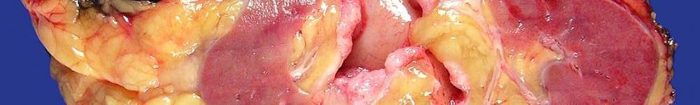
- Coronally bisect uterus through 3:00 and 9:00 positions.
- Measure endocervical canal and endometrial cavity in 2 dimensions, and thickness of endometrium and myometrium.
- Measure any lesions (whorled nodules, polyps, etc).
- Measure placenta and cord if present. Describe location of placental attachment to cavity.
Gross
- If any placenta is present, take representative umbilical cord (2 sections) and membrane roll as you normally would for a placenta.
- For placenta previs, look for placental tissue overlying the cervix and submit representative sections.
- For placenta accreta/increta/percreta, look for tissue adherent to the endometrium. Submit representative sections of the deepest extent of placental invasion into myometrium.
- If attached bladder peritoneum is present, be sure to sample this in continuity with outer myometrium/serosa.
- Gross the reminder of the specimen as a benign uterus.
Updated 6-22-2020 SRR
