Auto text: “Insert Trachelectomy”
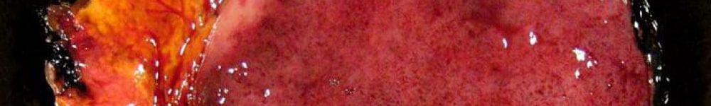
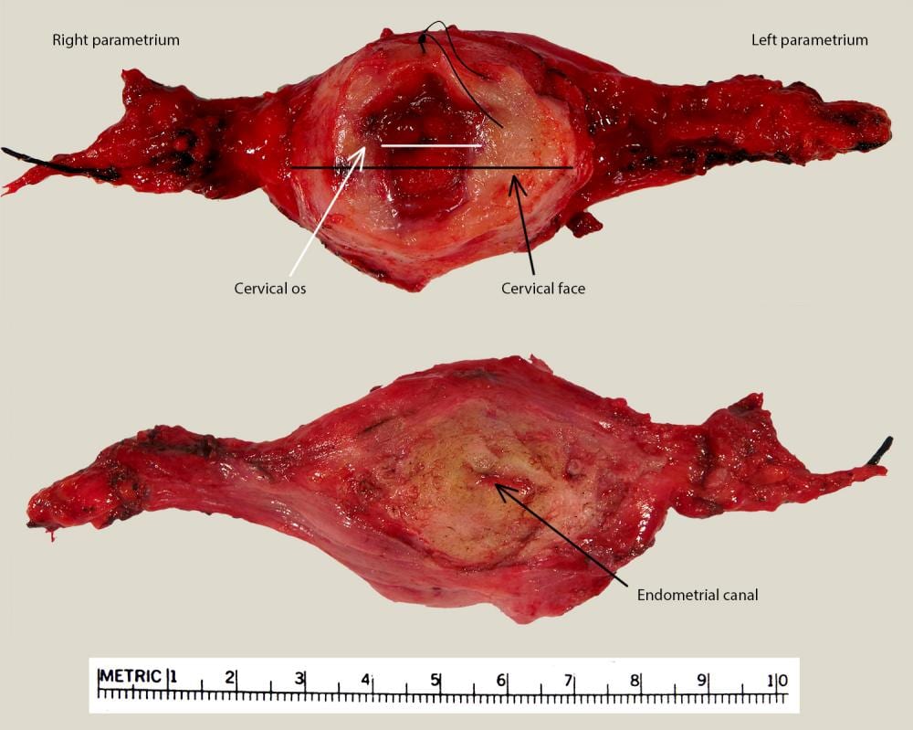
This specimen consists of a cervix with attached paracervical/parametrial tissue and a vaginal cuff. The procedure is performed to preserve fertility in women with early cervical cancer.
Triage
- Note any provided orientation. Usually there is a suture marking 12:00.
- Weigh specimen and measure:
- Overall dimension (3D).
- Cervical face (2D) – if prior LEEP/cone was performed, the cervical face may be distorted or absent.
- Shape and diameter of os.
- Width of vaginal cuff (give a range if variable).
- Right and left parametrial tissue (base x height of triangle).
- If a lesion is identified on the cervical face, photograph specimen prior to opening.

- Ink in 4 colors:
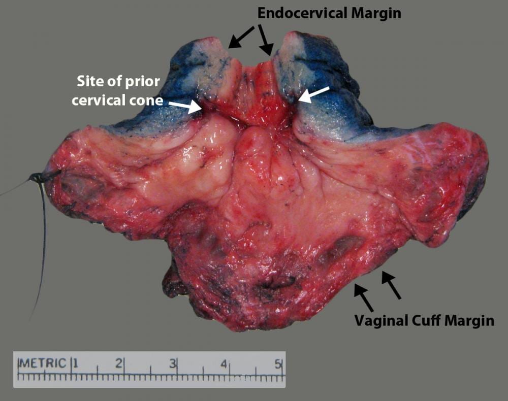
- Endocervical margin: Yellow.
- Anterior peripheral cervix and parametrium (9-3:00): Blue.
- Posterior peripheral cervix and parametrium (3-9:00): Black.
- Vaginal cuff: Red.
- Open cervix at 12:00, unless the carcinoma is clearly present at 12:00.
- Measure length of endocervical canal.
- Accurately measure lesion and note location. Measure it’s distance to the vaginal and endocervical margin.
- Photograph opened specimen with lesion.
- Pin on wax and fix in formalin.

Gross
- Serially section parametrial tissue in parasagittal plane and submit entirely from medial to lateral, noting right and left sides.
- Vaginal Cuff
- If no tumor is grossly present or measures <2 cm, submit the entire vaginal cuff margin in continuity with the ecto/endocervix in quadrants, making sure all sections include red ink (vaginal cuff).
- If tumor is grossly present and measures >2 cm and close to the vaginal cuff margin, submit sections perpendicular to margin, making sure your sections include ink. The remainder of the cuff margin can be trimmed and submitted en face in quadrants.
- If tumor is grossly present and measures >2 cm and far from the vaginal cuff margin (i.e. tumor and margin won’t fit in one block), the margin can be trimmed and submitted en face in quadrants.

- Serially section the tumor/specimen using full-thickness radial sections around the cervix.
- Measure depth of invasion, tumor thickness and distance from nearest inked deep margin.
- IF THERE IS NO VISIBLE LESION or tumor measures <2 cm, submit the entire specimen, radially around cervix.
- If tumor measures > 2 cm, submit representative sections of the lesion.
Updated 6-8-2022 SRR
