Auto text: “Insert Lung Explant”
- Weigh and measure (3D).
- Photograph intact specimen.

- Submit bronchial and vascular margins.
- Take 3-4 representative sections of hilar lymph nodes.
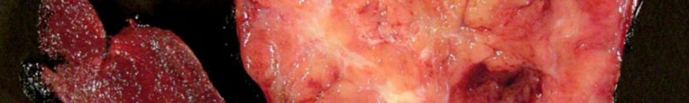
- Serially section sagittally and describe pleura, parenchyma, and airways.
- Photograph cut section.
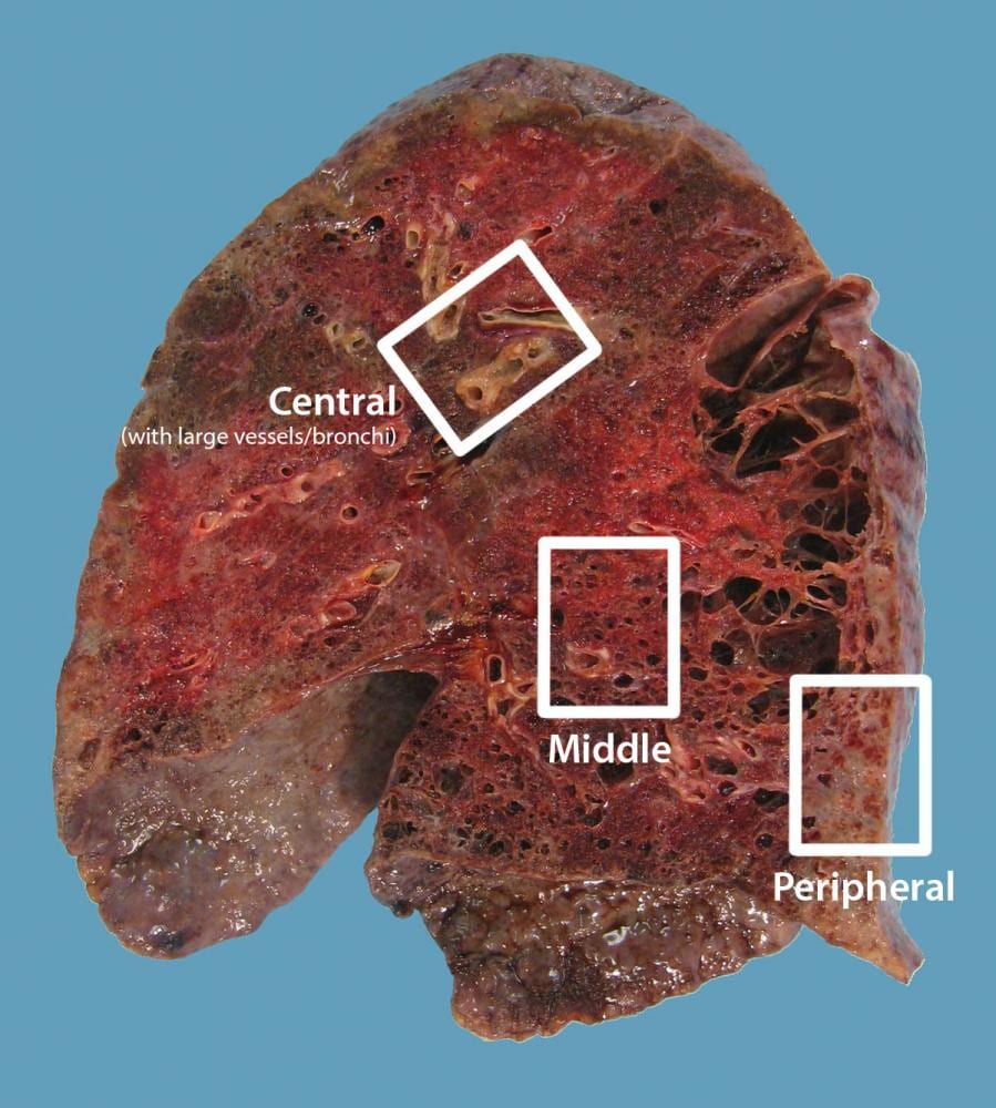
- Submit 3 sections per lobe for standard histology processing (central (hilar), middle, and peripheral; designate in block summary).


Updated 7/3/23 SRR
