Auto Text: “Insert Gallbladder”
Gallbladders are most often removed for cholecystitis or for cholelithiasis. On rare occasions, carcinoma is identified on gross exam. Pay attention for cancer in patients age 50 and over and submit 3 extra sections (see below).
Triage
- If there is a clinical or gross suspicion of malignancy, triage as follows:
- Ink the roughened outer surface of the gallbladder, which represents the area that abuts the liver.
- Obtain a cystic duct margin (en face) and save in cassette.
- Open specimen and fix in formalin.
- If non-neoplastic, the specimen can be cut fresh.
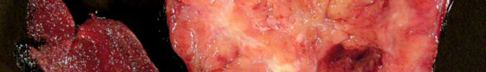
Gross
- If the gallbladder is received opened, that fact should be mentioned in the gross description.
- If for tumor, gross as follows:
- Measure in three dimensions.
- Obtain cystic duct margin (en face) if not already done.
- Measure mass (if identifiable) or range of wall thickness. Serially section through mass, noting depth of invasion and closest extent to inked margin.
- Submit the following:
- cystic duct margin (en face)
- multiple sections of tumor (7 cassettes), including closest to inked margin
- any polyps entirely
- representative normal gallbladder (2 cassettes)
- “sentinel” lymph node near cystic duct (and any other lymph nodes present)
- If benign, gross as follows:
- Measure in three dimensions.
- Remove the staple, which is closing the cystic duct – take a complete cross section of the cystic duct at the margin. Ink the periphery of the cystic duct to differentiate it from other representative sections.
- Describe the color and texture of the serosa.
- Open the specimen and measure the wall thickness. Describe the mucosa (velvety, firm, textured, discolored, etc).
- Describe the number, size, color, texture of any gallstones.
- Describe the amount, consistency and color of the bile.
- If otherwise normal, take a full length section of the gallbladder (from the fundus to the end of the cystic duct) and divide as necessary to fit into a cassette. DO NOT SUBMIT A “ROLL”. Include the separately taken cystic duct margin in the cassette as well.
- For all patients age 50 and over, take 3 additional (representative) sections in 1 cassette, even if no gross lesion is apparent.
- If any polyps are identified, submit them entirely. Also check the container for any “debris” that has a similar appearance to the polyp and submit that as well.
- A lymph node is usually present near the cystic duct (“sentinel node”). It should be submitted.
