Auto text: “Insert Uterus Lynch Syndrome”
Triage
- Weigh specimen and measure:
- 3 dimensions of uterus (C-C, Fundus-LUS, A-P).
- 3 dimensions of cervix (face and length).
- Shape, diameter of os.
- Ovaries (3D) and fallopian tubes (2D), if present.
- Identify anterior and posterior sides and note quality of serosa:
- The peritoneal reflection extends further inferiorly on the posterior side and is pointed.
- The peritoneal reflection on the anterior side is rounded.
- The tube is anterior to the ovary.
- Bisect uterus through 3:00 and 9:00 positions.
- Measure endocervical canal and endometrial cavity in 2 dimensions, and thickness of endometrium and myometrium.
- Measure any lesions (whorled nodules, polyps, etc).
- Most of these cases should be grossed on same day of receipt.
Gross
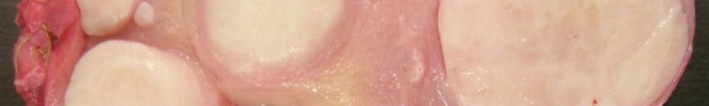
- Obtain longitudinal sections through cervix, anterior and posterior sides.
- Transversely section the endomyometrium (including the LUS) and take 4-6 full-thickness sections of (1 anterior LUS, 1 posterior LUS, 2 anterior endomyometrium and 2 posterior endomyometrium).
- Submit the remaining LUS, endometrium, and inner myometrium from “LUS” to fundus, maintaining orientation.
- Multiple sections can be placed in a single cassette.
- Submit any additional pathology (leiomyomas, polyps in their entirety, etc).
- Submit entire ovaries and fallopian tubes per SEE-FIM protocol.
- Amputate the distal 2 cm (fimbriae) and section parallel to the long axis.
- Section the remainder of tube transversely in 2-3 mm intervals.
- Adnexal soft tissue does not need to be submitted.
Updated 6-7-2022 SRR
