Auto text: “Insert Radical Nephrectomy”
Triage
- Check radiology reports for presence of tumor in renal vein and for number of arteries at hilum.
- Weigh and measure specimen in 3 dimensions.
- Measure length and diameter of attached ureter.
- Take margins (VERIFY ANATOMY AND SECTIONS WITH PA AS NEEDED):
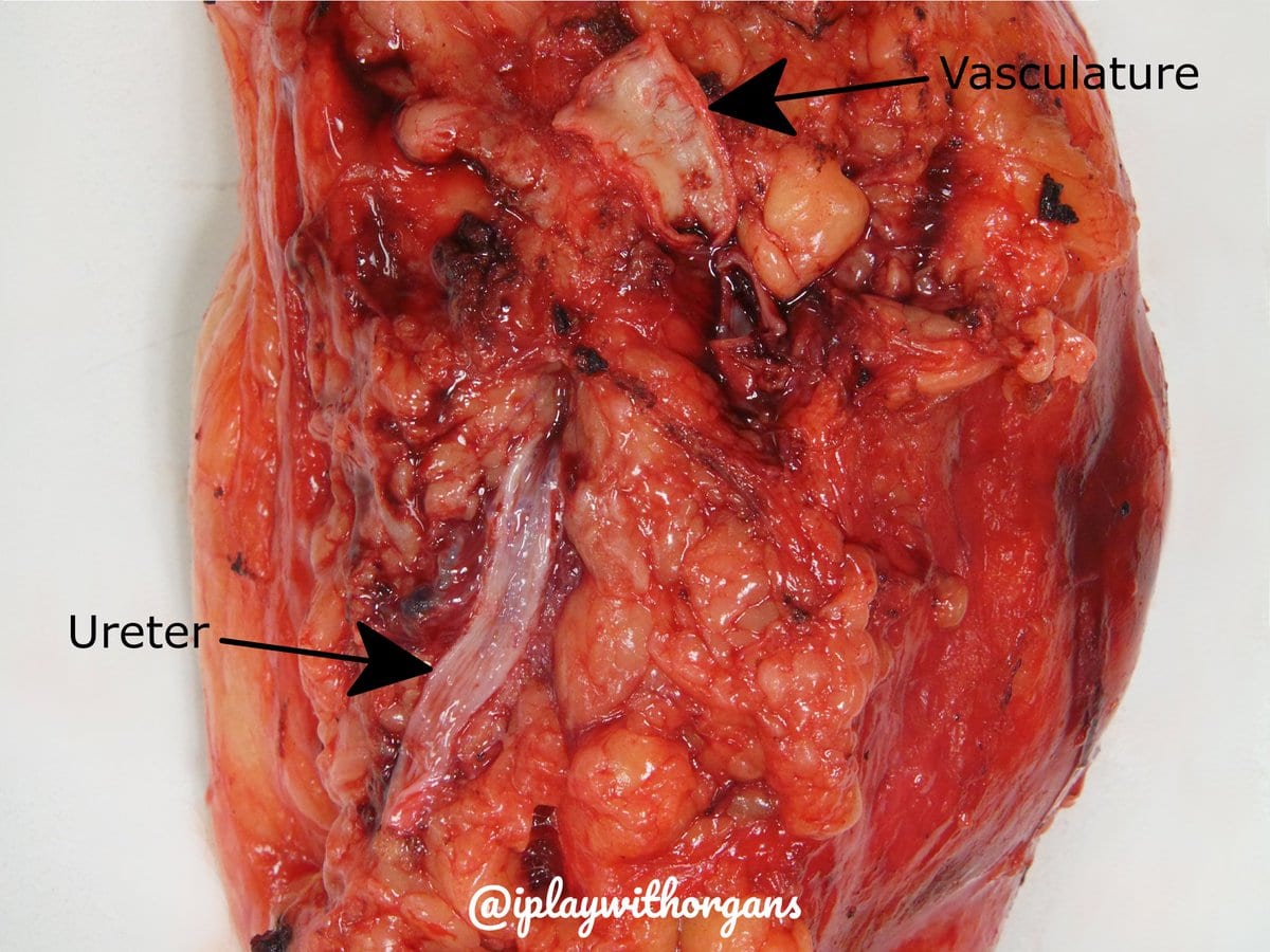
- Distal ureter margin, en face.
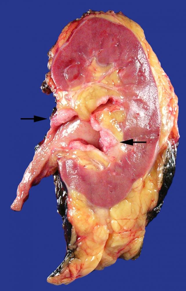
- Renal vessels at hilum, en face (artery/arteries and vein).
- Save in cassette within container.
- Tip from @iplaywithorgans: “Having trouble telling whether you have the ureter margin or just a vascular margin? Open them up. Ureters will have longitudinal corrugated lines, and vasculature will have a smooth lining”

- Ink outer surface in one color.
- Examine the renal vein for tumor thrombus. If present, note if it is adherent to the intima or not. Renal vein margin is considered positive ONLY if tumor thrombus has been transected and is attached to the wall of the renal vein at the transected surface. Margin is NOT considered positive if tumor thrombus has a smooth surface that bulges to the end of the vein.
- Open ureter and bisect the kidney kidney through renal pelvis/hilum.
- Identify and measure adrenal, if present.
- Identify and measure kidney, including cortical thickness.
- Identify and measure tumor (3 dimensions).
- Photograph cut section of kidney with tumor.
- If kidney is large, consider serially sectioning each half perpendicular to the long axis to aid fixation.
- Fix in formalin.
Gross
- Serially section each half of the kidney perpendicular to the long axis.
- Examine and describe:
- encapsulation of mass vs. circumcision vs. ill-defined
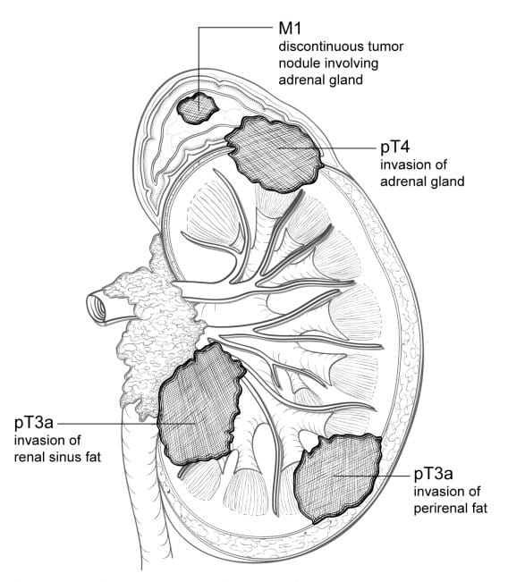
- invasion of tumor into perinephric or renal sinus adipose tissue
- invasion of tumor into renal vein, calyces, pelvis, ureter
- extension of tumor to inked surface
- non-neoplastic kidney parenchyma and cortical thickness
-

https://documents.cap.org/protocols/cp-kidney-17protocol-4011.pdf
- Look for lymph nodes within hilar and peripheral fat.
- Submit sections as follows:
- Ureteral and vascular margins (saved from triage).
- Tumor (1 per cm) including grossly-different areas and tumor with adjacent renal parenchyma.
- Deepest penetration of tumor into perinephric or renal sinus adipose tissue.
- Deepest penetration into renal vein.
- Tumor closest to the inked soft tissue margin (Gerota’s fascia).
- Normal cortex (add PAS-nephrectomy stain to this block).
- Any identified lymph nodes.
Renal Cell Carcinoma:

Urothelial Carcinoma: